
Unlocking the Secrets of Extra Fingers: A New Map for Ulnar Polydactyly
Alright, let’s dive into something pretty fascinating – those times when a hand decides it needs a little extra help, specifically on the pinky side. We’re talking about ulnar polydactyly, a congenital hand anomaly that pops up quite often. Now, you might think, “Okay, an extra finger, got it.” But hold on, it’s not always that simple! This condition can show up in a bunch of different ways, from just a tiny bump of skin to a fully formed extra digit.
For ages, folks in the medical world have tried to classify these variations to make sense of them, help with diagnosis, and plan the best way to fix things. Several systems have been proposed over the years, which is great, but honestly, they had a few blind spots. Some of the rarer ways ulnar polydactyly can manifest just didn’t fit neatly into the existing boxes. This made things a bit tricky when you encountered one of these less common presentations.
So, we got down to business. We decided it was time to create a classification system that could handle *all* the variations we were seeing, including those tricky, rare ones that slipped through the cracks of older systems. Our goal was to build something comprehensive, intuitive, and genuinely helpful for surgeons and clinicians dealing with these cases.
Why a New System? Filling the Gaps
You see, the existing classifications, while useful to a point, weren’t perfect. They sometimes lumped really different conditions together or just plain missed some specific types. For instance, we noticed cases where the duplication was just at the very tip of the finger (the distal phalanx) or, even more unusual, where an extra digit seemed to sprout from a deformed bone higher up in the hand (the fourth metacarpal). Previous systems didn’t have clear categories for these.
This lack of detail can make a real difference. If your classification system doesn’t accurately describe what you’re seeing, it’s harder to communicate effectively with other doctors, predict potential associated issues, and plan the most appropriate surgical approach. We needed a framework that was detailed enough to capture the full spectrum of ulnar polydactyly.
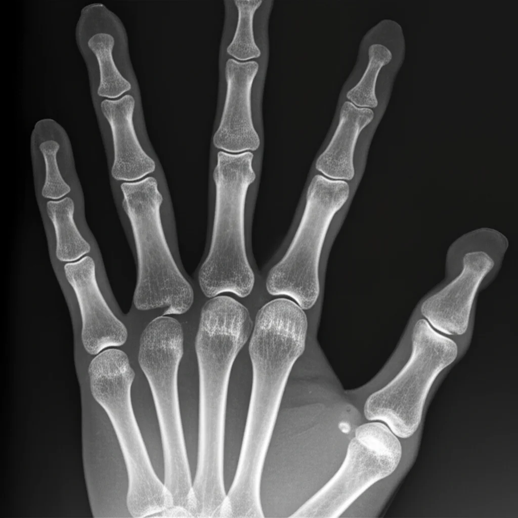
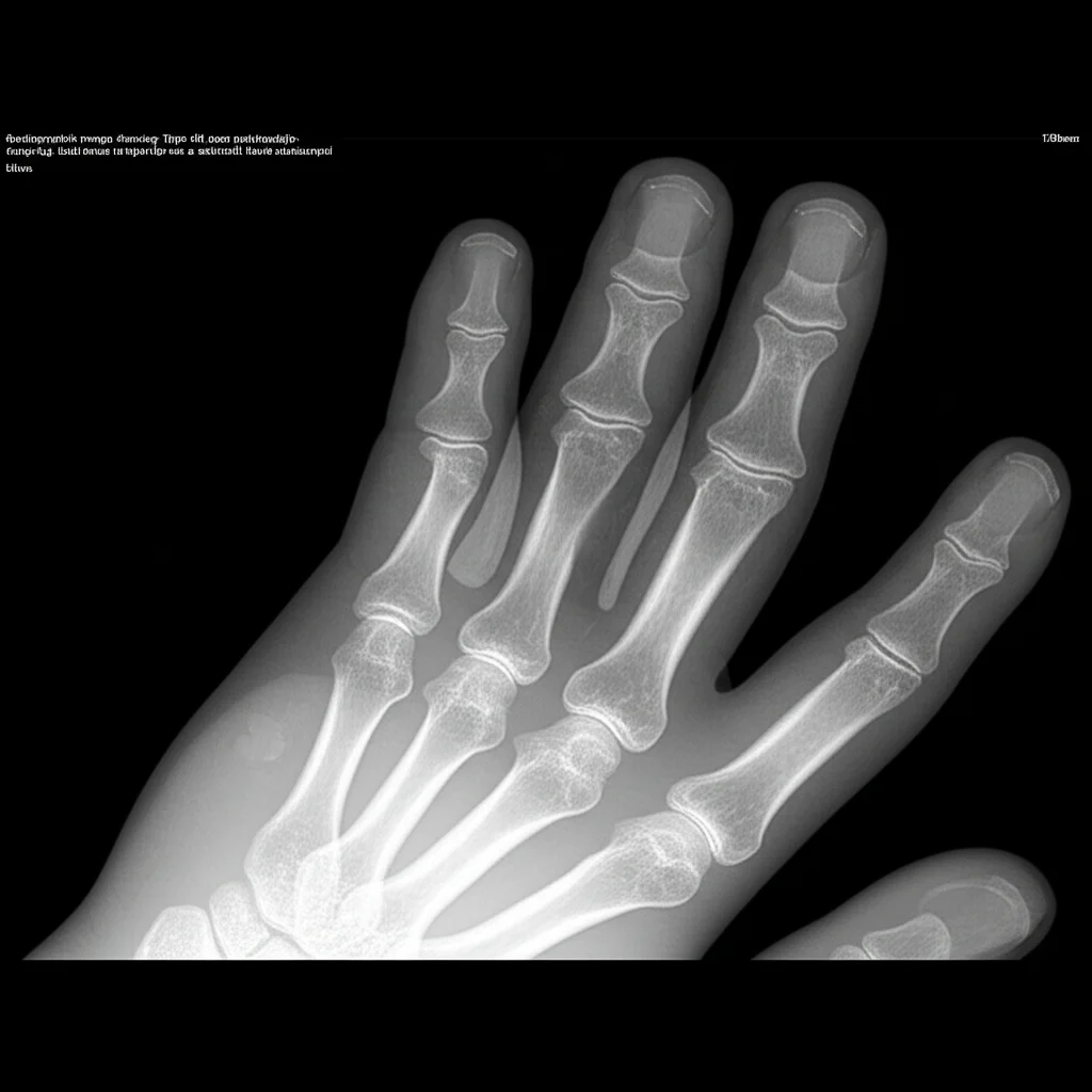
Our Approach: Getting Down to the Bones
To build our new system, we took a good look back at the cases we’d handled. We retrospectively reviewed the records of 35 patients with ulnar polydactyly who came through our doors between 2010 and 2022. We gathered all the juicy details: who they were, what their hands looked like, what the X-rays showed, if anyone else in the family had similar issues, if they had other health conditions, and what surgeries were done.
We poured over this data, looking at the morphology (what it looked like) and the radiographic findings (what the bones were doing). We also studied the existing classification systems, both for ulnar and radial (thumb side) polydactyly, particularly admiring the simplicity and logic of the Wassel classification used for radial types. We decided to base our system on the *most proximal level of skeletal involvement*. Think of it as tracing the duplication back towards the wrist – where does the extra bone (or lack thereof) start connecting or affecting the main hand structure?
After a lot of work and refinement, we landed on a system with five main types, numbered 0 through 4, and several subtypes within those. To make sure we weren’t just creating something that made sense to *us*, we tested its reliability. We had three independent hand surgeons classify a random set of cases using our new system, twice, with a couple of weeks in between. The results? Excellent agreement within each surgeon’s classifications (intra-observer reliability) and substantial agreement between different surgeons (inter-observer reliability). That told us our system was consistent and could be reliably applied by different people.
The Lowdown on Our Classification
Here’s the scoop on how we broke it down. Our system progresses from the simplest forms to the most complex, based on that proximal skeletal involvement we talked about:
- Type 0: Rudimentary Digits
These are the simplest forms, where the extra digit is underdeveloped and doesn’t have a bony connection to the hand.- Type 0a: Just a nubbin, maybe with a tiny nail.
- Type 0b: A floating digit connected by a skin bridge.
- Type 0c: A hypoplastic (underdeveloped) little finger that looks somewhat normal but isn’t connected by bone on X-ray (though we found a cartilaginous connection is usually there).
- Type 1: Distal Phalanx Type
Duplication or deformity involves the distal phalanx (the fingertip bone).- Type 1a: Bifid or irregular distal phalanx.
- Type 1b: A completely duplicated distal phalanx. (This was one of those rare ones not well-covered before!)
- Type 2: Middle Phalanx Type
Duplication or deformity involves the middle phalanx.- Type 2a: Bifid or irregular middle phalanx.
- Type 2b: Duplicated middle phalanx.
- Type 3: Proximal Phalanx Type
Duplication or deformity involves the proximal phalanx (the bone closest to the palm).- Type 3a: Bifid or irregular proximal phalanx.
- Type 3b: Duplicated proximal phalanx.
- Type 4: Metacarpal Type
Duplication or deformity involves the metacarpal bone (the long bone in the palm leading to the finger).- Type 4a: Bifid or irregular fifth metacarpal.
- Type 4b: A fully developed extra metacarpal articulating separately with the wrist bones.
- Type 4c: A duplicated, hypoplastic (underdeveloped) metacarpal.
- Type 4d: Duplication originating from a deformed *fourth* metacarpal. (Another rare one we specifically included!)
In our clinical series of 64 affected hands (one patient had surgery elsewhere, so that hand’s data was incomplete), Type 0 was indeed the most common, making up 38 cases (mostly 0b and 0c). Type 4 was the next most frequent with 19 cases (mostly 4a and 4b). Types 1 and 3 were rarer, with 3 and 4 cases respectively. Importantly, our system successfully categorized all cases, including those rare Type 1b and Type 4d presentations that were difficult to fit into older systems.
We also saw that having ulnar polydactyly often wasn’t the only thing going on. Thirty of our patients had other congenital conditions. Polydactyly of the foot was super common (22 cases), as was syndactyly (webbed fingers or toes, 15 cases). We also saw connections to radial polydactyly, triphalangism, club feet, and even conditions affecting the heart or brain. This highlights that these hand anomalies can sometimes be part of a larger picture or syndrome, like the one patient diagnosed with Greig cephalopolysyndactyly syndrome due to a specific gene mutation.
More Than Just Labels: Guiding Treatment
One of the coolest things about having a clear classification is how it helps guide treatment. Knowing the specific type tells us a lot about the underlying anatomy and potential challenges.
For the simpler Type 0 cases (the nubbins or skin-bridge digits), removal is usually straightforward. However, even here, our experience tells us that surgical excision is often better than just tying off the extra digit, as ligation can lead to more complications down the road. For Type 0c, where there’s no bone but often cartilage, we know we need to make sure that cartilaginous connection is fully removed to prevent it from growing back or causing issues.
When we get into Types 1, 2, and 3, where the bones of the finger itself are duplicated or deformed, the classification alerts us to potential complexities. We know to look closely at the nail bed (which might need careful management) and the finger joints (PIP and DIP joints), which might require reconstruction to ensure good function. Tendon reconstruction might also be necessary depending on how the muscles and tendons are attached to the duplicated parts.
Type 4, involving the metacarpal, often presents different options. Sometimes, you can preserve the whole extra finger if it’s well-formed, or you might need a more complex reconstruction, perhaps combining parts of the extra and main little fingers to create one well-functioning digit. Our classification prompts surgeons to think about the stability of the joint connecting the finger to the hand (the MCP joint) and whether ligaments and other structures need to be reconstructed to prevent the hand from spreading abnormally (metacarpal splay). If the bones are bifid or irregular (Types 1a, 2a, 3a, 4a, 4d), bone cuts (osteotomies) might be needed to properly align and separate the parts.
Looking Ahead: What’s Next?
Now, every study has its limits, and ours is no different. This was a look back at past cases from just one center, so the number of patients is finite, and we didn’t see examples of every single proposed subtype (like 1a, 2a, 2b). We included them in the system because the logic suggests they *could* exist based on the pattern of duplication, and we hope future studies with more cases will find them.
Also, because we were looking retrospectively, we couldn’t directly test if using our classification *predicted* better surgical outcomes or long-term function compared to using older systems. That’s definitely something we want to do in the future – follow patients prospectively to see how the classification helps guide decisions and improves results. We also didn’t do a deep dive into the post-operative outcomes in this particular study, which is another area for future research.
Despite these limitations, we really feel this new classification system is a big step forward. By specifically including previously unaddressed rare variants like the duplicated distal phalanx (Type 1b) and the duplication from the deformed fourth metacarpal (Type 4d), we’ve created a more complete picture of ulnar polydactyly. Organizing it based on the proximal skeletal level makes it intuitive and practical for surgeons, much like the Wassel system is for radial polydactyly. Plus, it gives us a way to systematically describe even complex cases by breaking them down into combinations of our basic types.
Ultimately, a better classification means a clearer understanding of the anomaly, more precise communication among clinicians, and hopefully, improved surgical planning and outcomes for kids born with these extra digits. It’s all about giving these hands the best possible start.
Source: Springer


