Unlocking Ferroptosis: How Toxicarioside H Takes Down Tough Breast Cancer Cells
Hey there! Let’s chat about something pretty important in the world of health – specifically, the fight against cancer. You know, breast cancer is a big deal globally, sadly ranking as the second leading cause of cancer-related deaths for women. It comes in different flavors, depending on certain markers, but there’s one type that’s particularly tricky: Triple-Negative Breast Cancer, or TNBC.
Why TNBC is Such a Challenge
Imagine a type of cancer that’s aggressive, likes to spread, and doesn’t respond to some of the usual targeted therapies because it lacks the common receptors (Estrogen, Progesterone, and HER2). That’s TNBC for you. It makes up about 15-20% of all breast cancers, and honestly, its prognosis isn’t the best. While surgery and chemo are standard, chemo often comes with tough side effects, resistance issues, and sometimes just isn’t effective enough, leaving the 5-year survival rate for advanced cases below 70%. So, yeah, finding new ways to tackle this beast is a pressing need.
Enter Cardiac Glycosides: More Than Just Heart Helpers?
Now, you might have heard of cardiac glycosides – they’re traditionally used for heart conditions. But guess what? Recent studies are showing they might have a hidden talent: fighting cancer cells! Our team has been busy isolating new ones from natural sources, and we stumbled upon a novel compound called Toxicarioside H (ToxH), found in the latex of a tropical plant. We’ve seen other cardiac glycosides from our work show promise against tumors through different mechanisms, like inducing autophagy or apoptosis. But the big question was: could ToxH trigger a specific type of cell death called ferroptosis in TNBC? That’s what we decided to dive into.
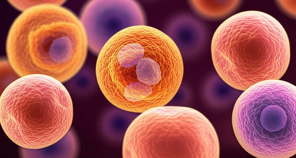
What We Found: ToxH Hits TNBC Hard
We started by testing ToxH on two specific TNBC cell lines, BT-549 and MDA-MB-468. Using standard lab tests like CCK-8 and EdU assays, we saw pretty clearly that ToxH put the brakes on their growth and proliferation. And it wasn’t just a little nudge; it was a significant, dose- and time-dependent smackdown. Higher doses and longer exposure meant less cancer cell activity. At concentrations between 50 and 250 µM, ToxH really started to inhibit growth, exceeding 50% inhibition at 200 and 250 µM. When we looked at 150 µM over time, the proliferation activity dropped significantly. To keep things manageable for later experiments focusing on *how* they die, we chose 100 µM and 150 µM for further study.
We also did EdU assays, which basically show us how many cells are actively dividing. Under the microscope and using flow cytometry, we saw a big drop in dividing cells after ToxH treatment. The 150 µM group had even fewer dividing cells than the 100 µM group. Pretty neat, right?
But here’s the really cool part: we also tested ToxH on normal breast epithelial cells (MCF-10A). And guess what? At the same concentrations that were hitting the TNBC cells, ToxH didn’t seem to bother the normal cells much at all. Their viability and proliferation were largely unaffected. This is super important because a good cancer treatment should ideally target cancer cells while leaving healthy ones alone.
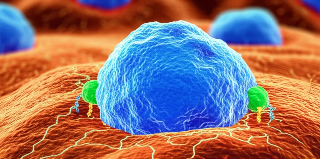
Unmasking the Death Dance: Is it Ferroptosis?
Cancer cells can die in different ways – apoptosis, autophagy, necrosis, and this newer one, ferroptosis. Ferroptosis is particularly interesting because it’s iron-dependent and involves a buildup of reactive oxygen species (ROS), leading to lipid peroxidation. It’s characterized by specific changes in mitochondria, like the cristae disappearing.
We used various methods to see how ToxH was killing the TNBC cells. Flow cytometry showed an increase in cells with damaged membranes (measured by 7-AAD staining) and changes in mitochondrial potential. The LDH release assay, which measures an enzyme released when cell membranes are leaky, also showed a significant release in ToxH-treated cells. This told us ToxH was definitely causing cell death by damaging the cell membrane. And again, the normal MCF-10A cells were mostly unaffected in these tests.
To figure out *which* type of death was dominant, we treated the TNBC cells with ToxH along with inhibitors for different death pathways: apoptosis (Z-VAD), necroptosis (Nec-1), autophagy (3-MA), and ferroptosis (Fer-1).
Transmission electron microscopy (TEM) images of ToxH-treated cells showed those characteristic ferroptosis signs – shrunken mitochondria with disappearing cristae – while the nucleus looked okay. This was a big clue!
When we looked at the percentage of dead cells (7-AAD positive) after adding the inhibitors, the ferroptosis inhibitor, Fer-1, was the champ at reducing ToxH-induced cell death. The LDH release results backed this up – Fer-1 was the most effective inhibitor in preventing the release of that leaky-membrane enzyme.
We also checked key markers of ferroptosis: lipid peroxidation (using BODIPY-C11) and free iron levels (Fe2+). ToxH treatment increased both of these, just as you’d expect in ferroptosis. Crucially, adding Fer-1 significantly *suppressed* both lipid peroxidation and Fe2+ generation caused by ToxH.

Putting all this together, it became clear: ToxH primarily inhibits TNBC cell growth by triggering ferroptosis. Fer-1’s ability to block ToxH’s effects really solidified this for us.
The Molecular Movers and Shakers: Nrf2 and HO-1
Okay, so ToxH causes ferroptosis. But *how*? We looked into the Nrf2/HO-1 pathway, which is known to be involved in iron metabolism and has been linked to ferroptosis. Nrf2 is a transcription factor that can influence genes related to ferroptosis, and HO-1 is an enzyme that breaks down heme, releasing iron.
Using Western blot analysis, we measured the levels of Nrf2 and HO-1 proteins in the TNBC cells after ToxH treatment. And bam! Both Nrf2 and HO-1 expression went up significantly, and this increase was dose-dependent – higher ToxH concentration meant higher Nrf2 and HO-1. This suggested that ToxH might be using this pathway to induce ferroptosis.
To really test if HO-1 was essential, we used a technique called shRNA to knock down, or silence, the HO-1 gene in the TNBC cells. Western blot confirmed that our shRNA successfully reduced both HO-1 and Nrf2 levels.
Then, we treated these cells (with silenced HO-1 or a control) with ToxH. Flow cytometry showed that when HO-1 was knocked down, the increase in dead cells (7-AAD positive) caused by ToxH was significantly reduced compared to the control group. The same thing happened with the ferroptosis markers: knocking down HO-1 significantly inhibited the increase in lipid peroxidation (BODIPY-C11) and free iron (Fe2+) that ToxH normally caused.
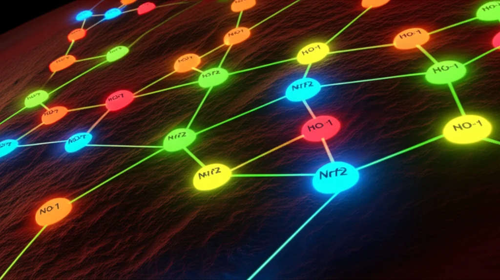
These results strongly suggest that HO-1 is a critical player, mediating the ferroptosis induced by ToxH in these TNBC cells. And since ToxH upregulates Nrf2, which in turn can affect HO-1, it seems the Nrf2/HO-1 pathway is indeed how ToxH triggers this specific cell death.
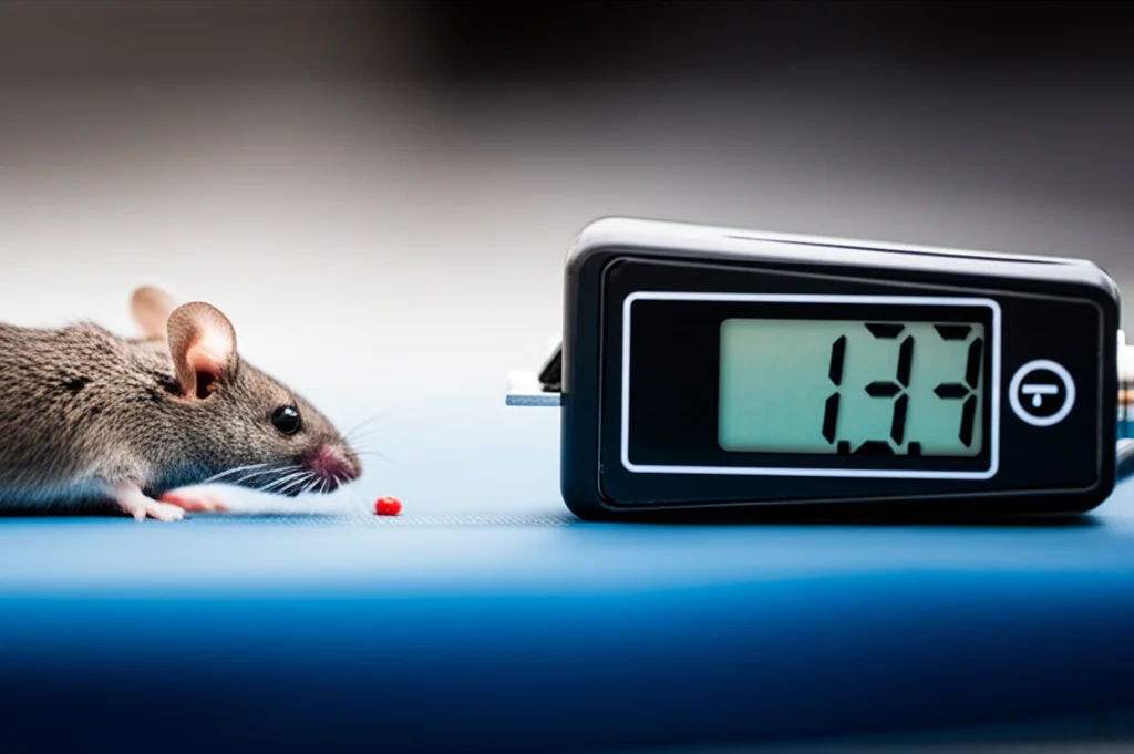
Taking the Fight to the Mice
Lab dishes are great, but what about a living system? We created mouse models with TNBC tumors using the same cell lines. We treated groups of mice with just a vehicle solution (the control), with ToxH, or with ToxH plus the ferroptosis inhibitor Fer-1.
Monitoring tumor volumes over 28 days, we were excited to see that ToxH treatment significantly suppressed tumor growth compared to the control group. It really put a dent in the tumor size! Interestingly, when we gave the mice both ToxH and Fer-1, the tumors were slightly larger than in the group that only got ToxH. This adds more evidence that ToxH is working by inducing ferroptosis – blocking ferroptosis partially counteracts ToxH’s anti-tumor effect.
We also kept a close eye on the mice’s overall health – body weight, lifespan, fur, eating, behavior. We didn’t see any significant negative impacts from the ToxH treatment compared to the control group. Examining their organs (heart, lungs, liver, spleen, kidneys) also showed no obvious signs of toxicity. This is fantastic news, suggesting ToxH might be effective against TNBC tumors *in vivo* with minimal side effects.
Finally, we checked the Nrf2 and HO-1 levels in the tumor tissues from the mice using Western blot. Just like in the lab dishes, ToxH treatment significantly increased the expression of both Nrf2 and HO-1 in the tumors. Adding Fer-1 didn’t seem to change these protein levels much compared to ToxH alone, suggesting ToxH’s effect on the pathway happens regardless of whether ferroptosis is blocked.
The Takeaway
So, what’s the big picture here? TNBC is a tough cancer, and we desperately need new treatment options. Our work shows that Toxicarioside H, a novel cardiac glycoside from a natural source, has some serious potential.
In the lab, ToxH effectively inhibits the growth and proliferation of TNBC cells and causes them to die, primarily through ferroptosis. It does this by revving up the Nrf2/HO-1 signaling pathway. And importantly, it seems to leave normal breast cells alone.
Taking it a step further, our experiments in mouse models confirmed that ToxH significantly suppresses TNBC tumor growth *in vivo*. The fact that blocking ferroptosis with Fer-1 partially reversed this effect in mice strengthens our conclusion about ToxH’s mechanism. Plus, ToxH appeared to be well-tolerated by the mice, which is a crucial factor for any potential therapy.
While we’ve uncovered a promising new angle – ToxH inducing ferroptosis via Nrf2/HO-1 in TNBC – there’s always more to learn. We need to dig deeper into the exact molecular steps involved. But these findings are super exciting! They position ToxH as a promising natural compound that could potentially be developed into a new treatment for TNBC. Further research, especially translating these findings towards clinical applications, is definitely warranted. It feels like we’re on the path to unlocking a powerful new weapon in the fight against this challenging disease!
Source: Springer


