Saving Little Feet: A Story of Hope After a Lawnmower Accident
Hey there. Let me tell you about something that really stuck with me – a story that shows just how incredible medicine and teamwork can be, especially when it comes to our little ones. We’re diving into a case about a brave 3-year-old boy and a truly nasty lawnmower injury. You see, while we might think of lawnmowers as just part of keeping the yard tidy, they can be incredibly dangerous, and sadly, kids sometimes get hurt. Injuries to the lower leg and foot are, unfortunately, the most common type when children are involved, making up a significant chunk of these accidents.
When the Unthinkable Happens: A Severe Injury
Now, most lawnmower injuries in kids, thankfully, aren’t *that* severe. They might involve cuts or scrapes that need some careful cleaning and maybe a special wound dressing system called VAC therapy (that’s Vacuum-Assisted Closure, a fancy way of saying it helps the wound heal by using suction). But sometimes, just sometimes, the injury is far more devastating. We’re talking major stuff, like losing part of a limb. These cases are rarer, but they require a whole different level of care – a team effort, really.
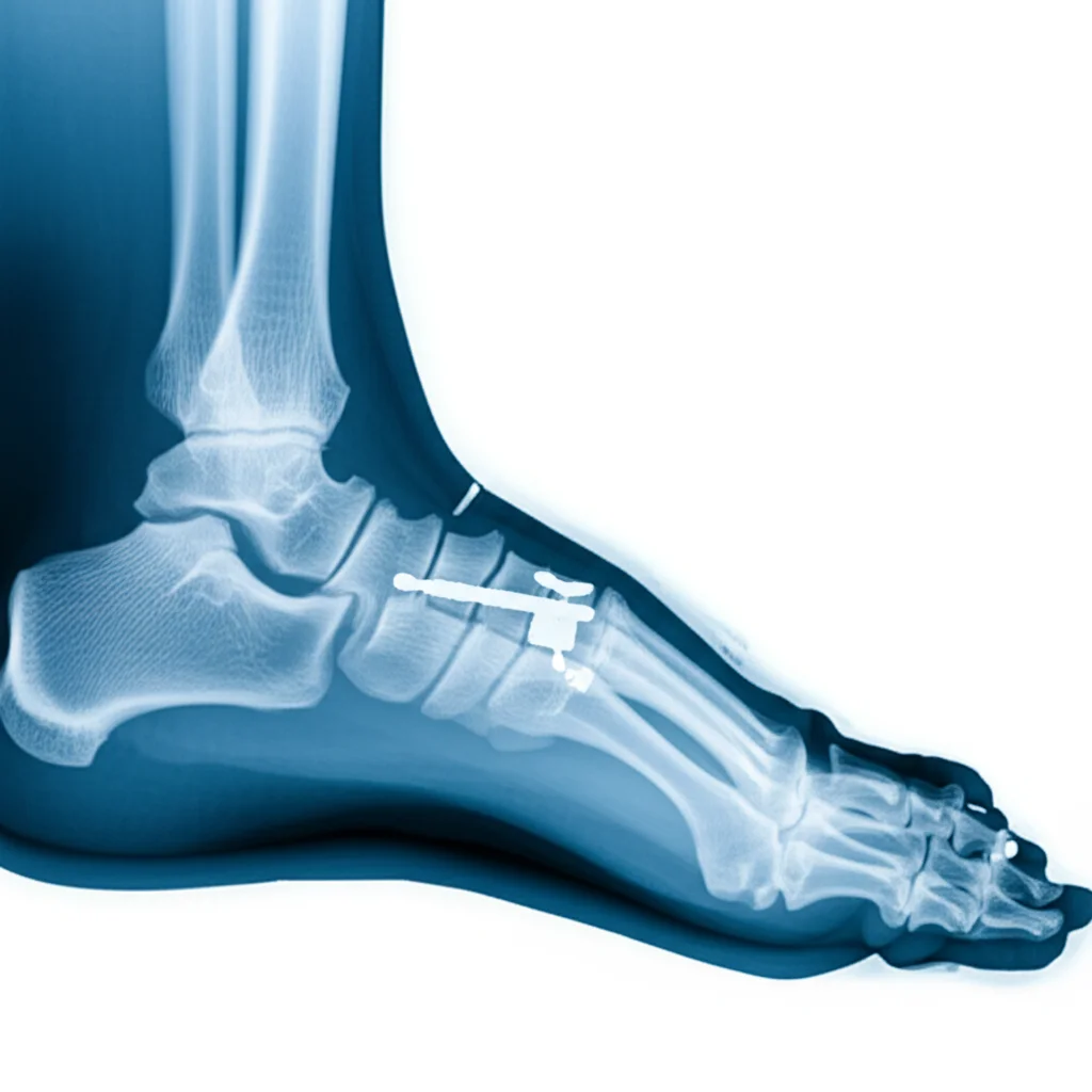
This particular story is about a little guy, just three years old, who had a terrible accident. A lawn tractor went over his left foot, causing a traumatic amputation right at the Chopart joint line. If you’re not familiar, that’s basically where the front part of the foot connects to the back part. Imagine that – the front of his foot was just gone. And if that wasn’t bad enough, the main bone in the back of the foot, the talus, had a big chunk missing, especially in the area where you bear weight. Looking at the initial X-ray, it was clear this was a massive injury.
The rescue team couldn’t even bring the amputated part to the hospital because it was just too damaged. When he arrived, his left foot was, frankly, destroyed. There was a huge open wound, tendons were visibly torn or cut, and those crucial bones in the front and middle of the foot were missing. The amputation was pretty much complete at that Chopart line, though a small piece of another bone, the cuboid, was partially still there. You could even see his foot starting to point downwards because the muscles that lift the foot were gone, and the strong Achilles tendon was pulling unopposed.
Given how severe and contaminated the wound was – lawnmowers pick up all sorts of dirt and debris – immediate reconstruction wasn’t an option. The first step was crucial: cleaning everything out thoroughly and starting that VAC therapy along with antibiotics to fight off infection. This VAC system was changed regularly, every few days, and it did its job, helping the foot start to heal and the wound bed to get ready for the next steps.
Preparing for the Big Fix
Identifying the damaged tendons was also key during this initial phase. They found stumps of some important ones – the tibialis posterior, and the flexor tendons that bend the toes. But the tendons that lift the foot and toes? They were nowhere to be found. This muscular imbalance was a major concern because, left untreated, it would definitely lead to a deformity where the foot points down, making it impossible to walk properly.
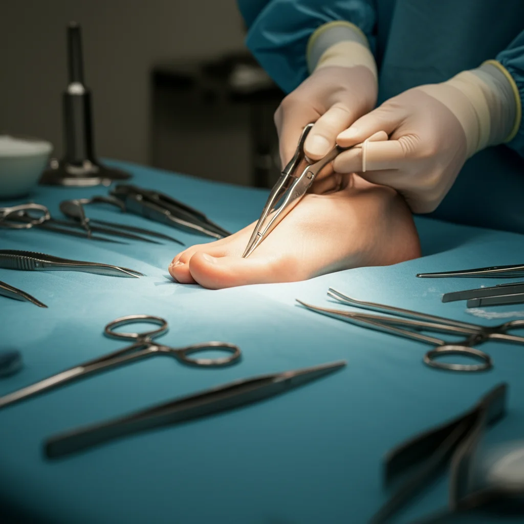
After several cycles of the VAC therapy, and once the wound was looking much healthier (about three weeks after the accident), it was time for the definitive surgery. This was the big one, requiring that multidisciplinary team I mentioned – orthopedic surgeons to deal with the bones and tendons, and plastic surgeons to cover the massive defect.
The main goal was twofold: fix the bone issue and restore some balance to the foot’s movement to prevent that dreaded equinus deformity. The orthopedic team started by addressing the Achilles tendon. They actually lengthened it using a technique called a Z-plasty. Imagine splitting the tendon lengthwise partway and then sliding the pieces past each other before sewing them back together – it makes the tendon longer. This was absolutely vital to counteract the pull that was making his foot point down.
Next, they worked on those remaining tendons. They cleverly rerouted some of the flexor tendons (the ones that bend things) and the peroneal tendons (on the side of the ankle) and attached them to the stumps of the missing extensor tendons (the ones that lift things). This is a bit like rewiring the foot to try and get some lifting function back and balance out the remaining muscles.
Then came the bone part. That small piece of the cuboid bone that was left? They reshaped it and fixed it to the damaged talus bone using special screws made of allogeneic bone (that’s bone from a donor, treated so the body accepts it). The idea here was to rebuild the weight-bearing surface of the talus, the part that takes the load when you stand and walk, using a piece of bone that still had its natural joint surface.
Rebuilding and Recovering
With the orthopedic work done, the plastic surgery team stepped in. Because the wound was so large, they needed a substantial amount of tissue to cover it. They chose a free latissimus dorsi flap – that’s a piece of muscle and skin from the back, which they carefully detached, including its blood vessels. They then brought this flap down to the foot and painstakingly connected its blood vessels to vessels in the ankle using microsurgery. This flap provided the necessary coverage and healthy tissue for the foot to heal. Finally, they covered the muscle flap with a thin layer of skin taken from the boy’s thigh.
After this marathon surgery, the foot was immobilized in a cast. Pain was managed with a nerve block, which is great because it provides continuous relief and can even help with blood flow to the flap. The leg was kept elevated to help the flap settle in. The cast stayed on for six weeks to protect everything – the tendon transfers, the bone fixation, and the healing flap.
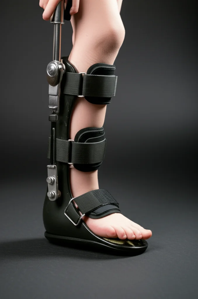
Once the cast came off, the next phase began: getting the foot used to bearing weight again. He was fitted with a special offloading orthosis, a brace that helped distribute the pressure and allowed him to start putting some weight on the heel, which was now the lowest point of his foot thanks to the Achilles lengthening. Physiotherapy was crucial throughout this period to help him regain function and adapt.
He used this orthosis for about four months, gradually becoming fully mobile without pain. After that, he transitioned to a different type of orthosis, an “OrthoPro,” which fits inside a normal shoe. This was a huge step towards getting back to a more typical childhood experience.
The Outcome: A Functional Foot and Preserved Quality of Life
Fast forward a year after that complex surgery, and the results are truly remarkable. The boy is doing great. He uses his orthosis perfectly and can bear full weight in his everyday life without limitations or pain. The Achilles lengthening did its job, ensuring his heel is the weight-bearing point. Crucially, there’s no significant difference in leg length, which is a massive win.
This outcome wasn’t guaranteed. A severe injury like this could very easily have resulted in a below-knee amputation. While that might seem like a simpler, one-step solution, especially given the complexity of reconstructing the foot, it comes with significant long-term consequences, particularly for a growing child.
Think about it: a below-knee amputation means a lifetime dependency on a prosthesis. While prosthetics are amazing, they can still limit certain activities and require constant adjustments as a child grows. It also results in a noticeable leg length discrepancy.
The approach taken here – a Chopart amputation level combined with complex reconstruction – aimed to preserve as much of the limb as possible, specifically the ankle joint and the heel. Why? Because preserving leg length and the ability to bear weight directly on the foot (even with an orthosis) dramatically improves quality of life. Imagine being able to get up in the middle of the night to use the bathroom without needing to put on a prosthesis first. Or the ability to just spontaneously run and play, which is so important for a young child.
Yes, this kind of reconstruction is complicated. Chopart amputations can have higher risks of wound healing problems compared to a straightforward below-knee amputation. But with a skilled, multidisciplinary team, careful wound management (like the VAC therapy that prepared the site so well), and meticulous surgical technique, these risks can be managed. The latissimus dorsi flap was a good choice here because the defect was so big, and it’s known for having reliable blood vessels, even in young kids. Plus, muscle flaps tend to shrink over time, which can lead to a better final shape without needing more surgeries later.
Dealing with young patients also presents challenges, like ensuring they cooperate with post-op care. Using a cast with a window for monitoring the flap and effective pain management (like that nerve block) were smart strategies to help with this.
The Takeaway: Teamwork Makes the Dream Work
This case really highlights the value of a multidisciplinary approach. Orthopedics, plastic surgery, physiotherapy, orthotics – everyone played a crucial role. It wasn’t about the fastest or simplest fix, but about the *best* long-term outcome for this little boy. They weighed the options: a quick amputation with lifelong dependency versus a complex series of procedures aimed at preserving function and independence.
For young patients with severe foot trauma, especially from something like a lawnmower, preserving leg length and aiming for a weight-bearing foot, even if it means more initial surgeries and ongoing orthotic support, can make a world of difference in their quality of life as they grow. It’s a testament to what can be achieved when specialists work together with a focus on the patient’s future.
Source: Springer