P2Y2R Unlocked: Peeking into its Secret Life of Self-Starts and Protein Pals!
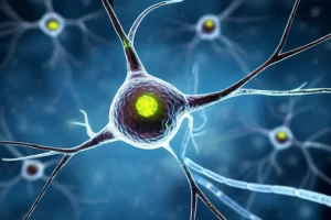
Hey there, science pals! Ever wonder how our cells chat with each other? It’s a super complex world down there, and one of the ways they do it is through tiny little messengers and the even tinier “mailboxes” that receive them. Today, I want to take you on a bit of a journey into the life of one such mailbox, a super important protein called the P2Y2 receptor (P2Y2R). It’s a real workhorse in our bodies, and we’ve just gotten some incredible new peeks into how it does its job. Buckle up, because it’s pretty cool stuff!
P2Y2R: Why’s This Tiny Protein Such a Big Shot?
So, what’s the big deal with P2Y2R? Well, it’s part of a family called purinergic receptors, which basically means they respond to molecules like ATP and UTP – you know, the energy currency of the cell, but turns out, they’re also big-time signaling molecules outside the cell! P2Y2R is like a sensor on the cell surface, waiting for these ATP or UTP signals. When it gets one, it kicks off a chain reaction inside the cell. This little guy is found all over the place: in our muscles, heart, lungs, gut, you name it. And it’s involved in a ton of important processes:
- Helping our blood vessels relax.
- Managing how fluids move in our airways (super important for breathing!).
- Guiding our immune cells to where they’re needed.
- Playing a role in wound healing.
- Even helping with the early development of heart tissue!
It does all this by “talking” to different partners inside the cell called G-proteins. Think of G-proteins as the next link in the communication chain. P2Y2R is known to chat with Gq/11 proteins, which usually leads to a calcium signal inside the cell. But here’s a fun twist: it can also cozy up to G12 and Go proteins, especially when it’s hanging out with other proteins called integrins. This can change how cells move and how their internal skeletons are arranged. Pretty versatile, huh?
Now, despite knowing P2Y2R is a big deal, understanding exactly how it recognizes its signals (we call them ligands) and how it chooses its G-protein partners has been a bit of a mystery. Why? Because we didn’t have a clear picture of what it actually looks like when it’s doing its thing. That’s where the real adventure begins!
Seeing is Believing: How We Got a Snapshot of P2Y2R in Action
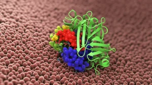
To really understand a machine, you need to see its parts, right? For proteins, which are nano-machines, this means getting a “structural” view. And that’s exactly what we set out to do! We used a super cool technique called cryo-electron microscopy (cryo-EM). Imagine taking thousands upon thousands of pictures of these tiny protein complexes, frozen in action, and then using powerful computers to combine them into a 3D model. It’s like molecular photography at an insane resolution!
We managed to get several snapshots:
- P2Y2R all by itself (we call this the ‘apo’ state) hooked up to a Gq protein.
- P2Y2R with ATP (one of its main activators) bound to it, also hooked up to either a Gq or a Go protein.
- And just for comparison, we also looked at a close cousin, P2Y4R, with UTP bound to it and hooked up to Gq.
These structures are like gold! They let us see, for the first time, the nitty-gritty details of how P2Y2R works. We could see exactly where ATP and UTP fit, how the receptor changes shape, and how it grabs onto its G-protein partners. It’s like finally getting the assembly instructions for a really complex piece of IKEA furniture, but way more exciting (and important for health!). The level of detail was amazing, down to how specific bits of the protein, called helices, move and twist.

One thing we noticed right away is that P2Y2R, like many similar receptors, has a classic seven-transmembrane helical bundle structure – imagine seven rods passing through the cell membrane. And there are these crucial ‘disulfide bonds’, like tiny molecular staples, holding parts of its extracellular loops in the right shape. We even saw some sterol-like molecules (think cholesterol relatives) snuggled into the receptor, probably helping to keep it stable. It’s these kinds of details that help us piece together the whole story.
The Handshake: How P2Y2R Recognizes Its Molecular Messengers
Alright, so P2Y2R is waiting for ATP or UTP. How does it actually “see” and “grab” them? Our structures gave us a front-row seat to this molecular handshake. The binding pocket for ATP in P2Y2R is relatively shallow and is formed by bits of those transmembrane helices, but also, very importantly, by its N-terminus (the very beginning of the protein chain) and a part called extracellular loop 2 (ECL2).
The N-terminus actually folds over like a little cap, helping to define the top of the pocket. And ECL2 dives down into the pocket. It’s a team effort! When ATP comes along, its negatively charged triphosphate tail (the “TP” part) nestles into the bottom of the pocket. It makes a whole bunch of connections with positively charged residues (amino acids) in the receptor, like K32, R110, and R292. It’s like a perfect magnetic fit. This is super important, because if you use ADP (which only has two phosphates), it doesn’t bind as well, and the receptor isn’t activated as strongly – something we also confirmed with our experiments!
The adenine base of ATP (the “A” part) gets sandwiched between two aromatic residues in that N-terminal cap, Y23 and F27, through what we call π–π interactions – think of it like molecular Velcro. The ribose sugar part of ATP also makes a specific hydrogen bond. It’s all incredibly precise!
Now, comparing P2Y2R with its cousin P2Y4R was really insightful. P2Y4R also binds UTP (which is very similar to ATP but has a uracil base instead of adenine) in a similar way, using many of the same contact points for the triphosphate tail. However, the N-terminus is a bit different. This difference in the N-terminus seems to be key to why P2Y2R is a full-on fan of ATP, while P2Y4R is a bit more lukewarm towards ATP (it prefers UTP). We even did some “swap” experiments, changing a few amino acids in the N-termini of P2Y2R and P2Y4R to make them more like each other. And guess what? P2Y4R with a P2Y2R-like N-terminus got better at responding to ATP, while P2Y2R with a P2Y4R-like N-terminus became less enthusiastic about ATP. Science is cool, right? This tells us that N-terminus is a real gatekeeper for ligand specificity.
It’s also interesting to compare these P2Y1R-like receptors (P2Y1R, P2Y2R, P2Y4R) with the P2Y12R-like family. The P2Y1R-likes have a shallower pocket where the ligand sits “base-up.” P2Y12R, on the other hand, has a deeper, more sealed pocket, and the ligand sits “base-down.” These are fundamental differences that explain why they respond to different molecules and why designing drugs for one doesn’t automatically work for the other.

A Social Butterfly: P2Y2R’s Flexible Friendships with G-proteins
Once P2Y2R grabs its ATP or UTP, it needs to pass the message along. It does this by coupling with G-proteins. We knew P2Y2R primarily couples to Gq/11 proteins, but our experiments showed it’s quite the social butterfly! It can also strongly engage with Gq, G12, G13, and importantly, Go proteins. It wasn’t so keen on Gs or the Gi subtypes.
This promiscuity is fascinating. Why would it want to talk to different G-proteins? Well, different G-proteins trigger different signaling pathways inside the cell, leading to different outcomes. For example, Gq often leads to calcium release, while Go can be involved in things like changing the cell’s shape or how it moves – super relevant for processes like wound healing or nerve cells growing extensions.
Our cryo-EM structures of P2Y2R coupled to Gq and P2Y2R coupled to Go were invaluable here. We could directly compare how the receptor interacts with these two different partners. The overall shape of P2Y2R was quite similar in both complexes, but there were subtle but crucial differences in how it “held hands” with Gq versus Go.
Choosing Favorites: The Secret Sauce Behind P2Y2R’s G-protein Picks
So, how does P2Y2R manage these different G-protein relationships? It’s all in the details of the interaction surface. The main docking site for the G-protein on the receptor is a cavity formed by several of those transmembrane helices (TM2, TM3, TM5, TM6, TM7) and the intracellular loops (ICLs) that connect them.
When we compared the P2Y2R-Gq structure with the P2Y2R-Go structure, we noticed that the G-protein itself (specifically its α5 helix, a key part for receptor binding) sits slightly differently. The α5 helix of Go is rotated a bit and shifted closer to TM3 and ICL2 of the receptor compared to Gq.
This shift allows for specific interactions. For instance:
- For Go coupling: A part of P2Y2R called ICL3 (intracellular loop 3) seemed to be really important. In the P2Y2R-Go structure, ICL3 was well-ordered and made several specific contacts with the Go protein. In the P2Y2R-Gq structure, ICL3 was more flexible and less defined. When we mutated key residues in ICL3, the receptor’s ability to activate Go was significantly hampered, more so than its ability to activate Gq. This suggests ICL3 plays a more dominant role in cozying up to Go.
- For Gq coupling: ICL2 seemed to be more critical. A residue L139 in ICL2 of P2Y2R snuggled into a hydrophobic pocket on Gq. While a similar interaction could happen with Go, it looked weaker because L139 was positioned a bit differently. Mutating L139 hit Gq activation harder than Go activation. Also, a residue H130 in TM3 made a stronger connection with Gq than with Go.
It’s like having different handgrips for different partners. These subtle structural variations on the intracellular side of P2Y2R allow it to differentiate between Gq and Go, leading to pathway-specific signaling. This is super important because it means the cell can fine-tune its response depending on which G-protein P2Y2R decides to activate more strongly. Understanding these specific “handgrips” could even give us ideas for designing drugs that encourage P2Y2R to pick one G-protein partner over another – talk about precision medicine!

The “It Does It Itself!” Trick: P2Y2R’s Built-in Starter Key
Now for perhaps the most surprising part of our P2Y2R story! We noticed that P2Y2R has a pretty high level of “constitutive activity.” This means it’s somewhat active even when there’s no ATP or UTP around to officially switch it on. This had been hinted at in previous studies, but we wanted to know why.
To investigate this, we looked at the structure of P2Y2R coupled to Gq, but this time without any ATP or UTP added (the ‘apo’ state). And what we saw was remarkable! A part of the receptor’s own N-terminus, a little helix-like segment from T15 to R24, was actually folded back and tucked right into the orthosteric ligand-binding pocket – the very same pocket where ATP normally binds!
It’s like the receptor has its own “built-in agonist” or a starter key. This N-terminal segment makes a bunch of specific interactions with the pocket, mimicking some of the interactions ATP would make. For example, T15 from the N-terminus hydrogen bonds with Y268 in TM6, a residue that’s also important for ATP binding. D17 from the N-terminus interacts with R272, another key player in ATP recognition.
To test if this N-terminal “key” was really responsible for the self-activation, we did a few things:
- We made mutants where we progressively replaced parts of this N-terminal helix with a simple linker. The more of the helix we replaced, the less constitutive activity the receptor had.
- We even synthesized a peptide corresponding to this N-terminal segment and found it could activate a P2Y2R that had its own N-terminus chopped off!
This is a super cool mechanism! It means P2Y2R isn’t just passively waiting for a signal; it has a way to keep itself partially “on” all the time. This self-activation is stabilized by that disulfide bond we mentioned earlier, C25 on the N-terminus connecting to C278 in TM7, which helps anchor the N-terminus in place. If we broke this bond, the constitutive activity pretty much vanished.
This “built-in agonist” idea isn’t entirely new in the world of GPCRs (the big family P2Y2R belongs to). Some orphan GPCRs (receptors whose natural activators are unknown) use similar tricks with their N-termini or extracellular loops to self-activate. But the way P2Y2R does it, with this relatively shallowly-docked N-terminal helix, is unique. And it seems this feature is conserved in mammals, suggesting it’s an important regulatory mechanism.
When this N-terminal segment is in the pocket, it causes the receptor to adopt an active-like shape, allowing it to couple to Gq. However, the interactions with Gq are a bit weaker compared to when ATP is bound. This makes sense – self-activation is like an idling engine, while ATP binding is like putting the pedal to the metal for full activation.

So, What’s the Big Picture?
Phew! That was a deep dive, wasn’t it? What we’ve learned about P2Y2R is pretty groundbreaking. We now have a much clearer picture of:
- How it recognizes ATP and UTP: The specific roles of the N-terminus and ECL2, and the key amino acid contacts. This is crucial for understanding why it prefers certain ligands.
- How it chooses its G-protein partners (Gq vs. Go): The subtle differences in the intracellular binding surface, particularly involving ICL2 and ICL3, give us clues about its signaling versatility.
- Its surprising self-activation mechanism: The N-terminus acting as a “built-in” agonist explains its constitutive activity and adds another layer to how its function is regulated.
Why does all this matter? Well, P2Y2R is involved in so many physiological processes, and it’s also implicated in diseases like dry eye syndrome, cystic fibrosis, chronic inflammation, and even some cancers. Understanding its structure and how it works at a molecular level is the first step towards designing better drugs.
For example, now that we see the details of the ATP binding pocket and the role of the N-terminus, we could try to design drugs that are more selective for P2Y2R over its cousins like P2Y4R. Or, knowing how it interacts differently with Gq and Go, perhaps we could develop “biased agonists” – drugs that nudge P2Y2R to activate one pathway more than another. And the discovery of the N-terminal self-activation pocket opens up a whole new avenue for designing peptides or small molecules that could modulate P2Y2R activity, perhaps by mimicking or blocking this self-starter key.
It’s a fantastic example of how basic science – just trying to understand how things work – can have real-world implications. These tiny molecular machines hold so many secrets, and every new structure, every new insight, brings us a little closer to harnessing their power for good. It’s a reminder that there’s a whole universe of complexity and elegance happening inside us all the time!
The research journey involved a lot of meticulous work, from preparing the proteins, collecting tons of cryo-EM data, to complex data processing and building the atomic models. Functional assays in cells helped confirm what the structures were telling us, and molecular dynamics simulations gave us a sense of how these proteins move and breathe. It’s always a team effort in science!
Source: Springer