Unlocking Epilepsy’s Secrets: Why New MRI Tech Beats the Old Guard
Alright, let’s talk about something pretty significant in the world of brain science and health: epilepsy, specifically the kind that hangs out in the temporal lobe. If you or someone you know deals with this, you know it’s no walk in the park. Idiopathic temporal lobe epilepsy (ITLE) is actually the most common type of focal epilepsy in adults, and honestly, it can be stubborn. Sometimes, medication just doesn’t cut it, and folks might need to consider surgery to get those seizures under control.
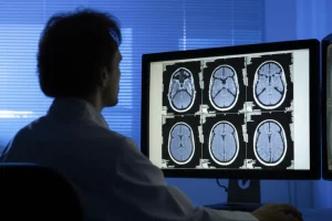
Now, figuring out exactly *where* in the brain these seizures are starting is crucial, especially if surgery is on the table. This is where MRI comes in – it’s been our go-to tool for peering inside the brain. Conventionally, doctors look at MRI scans visually, checking for tell-tale signs like changes in the hippocampus, that seahorse-shaped structure deep inside your temporal lobe that’s super important for memory and often involved in TLE.
But here’s the kicker: about 30% of people with TLE don’t show any obvious problems on these standard visual MRI scans. We call these “MRI-negative” cases, and they’re a real puzzle when you’re trying to pinpoint the seizure source. This got us thinking – could there be a better way?
Enter the Quantitative Heroes
This is where our story gets interesting. Beyond just looking at pictures, we can use quantitative methods with MRI. Think of it like moving from just glancing at a photo to actually measuring everything in it with precision tools. Two techniques have shown real promise:
- Automated MR Volumetry: Instead of just eyeballing if a structure looks smaller, this uses software to automatically measure the exact volume of brain regions like the hippocampus and amygdala. It’s objective, consistent, and can spot subtle differences that might be missed visually.
- Proton MR Spectroscopy (MRS): This isn’t about structure; it’s about chemistry! MRS looks at the levels of different metabolites in the brain tissue. Metabolic changes can happen *before* structural damage is visible, potentially making it an earlier and more sensitive detector of problems.
So, we set out on a mission. Our objective was clear: let’s see if these quantitative techniques – automated MR volumetry and MRS – could do a better job than conventional visual MRI at two key things in idiopathic TLE:
- Lateralization: Pinpointing which side of the brain (left or right) the seizures are coming from.
- Prognostic Assessment: Getting clues about how the epilepsy might behave, perhaps predicting who might have a tougher time controlling seizures with medication.
Our Deep Dive into the Brain
We put together a study involving 33 patients with idiopathic TLE and 40 healthy folks who were similar in age and sex to act as our control group. The patients were already classified into right or left TLE based on their EEG results (that’s the test that measures electrical activity in the brain).
Everyone got a good old history-taking and clinical check-up. Both groups also had standard MRI and MRS scans. The patient group, because they were the ones we were really studying, also had EEG and automated MR volumetry done. Then came the fun part – comparing what each of these MRI techniques told us.
We wanted to see which one was best at finding the seizure focus and which could give us hints about how well the epilepsy was controlled. We used a 1.5-T MRI machine, which is pretty standard, and followed a specific protocol to get the best images, including special sequences to look closely at the temporal lobe and hippocampus.
For the automated volumetry, we took the 3D MRI scans and fed them into a cool online platform called volBrain. This platform automatically segments the brain and gives precise volume measurements for different structures, like the hippocampus and amygdala. It even provides normal ranges based on age and sex and calculates asymmetry indices – basically, how different one side is from the other. This is way more objective than just looking!

For MRS, we placed a small “voxel” (think of it as a tiny box) in the temporal lobe, specifically including part of the hippocampus. MRS then measures the levels of key brain chemicals like NAA (N-acetylaspartate, often seen as a marker of neuronal health), Cr (Creatine, involved in energy), and Cho (Choline, related to cell membranes). We calculated ratios like NAA/Cr, Cho/Cr, and NAA/(Cho+Cr) because ratios are often more reliable than absolute values. Changes in these ratios can indicate neuronal dysfunction or damage. We also used an asymmetry index for MRS, comparing the ratios on the left and right sides.
Conventional MRI scans were reviewed by two experienced radiologists who didn’t know anything about the patients’ clinical history or EEG results – this helped keep things unbiased. They looked for changes in signal intensity (how bright or dark areas appeared) and visible volume loss (atrophy) in the temporal lobe structures.
The Results Are In!
So, what did we discover when we crunched all the numbers?
First, let’s talk about lateralization – figuring out which side is the problem. This is arguably the most critical piece of information for potential surgery.
Conventional visual MRI, looking for signal changes and atrophy, could only confidently lateralize the seizure focus in about a third of our patients (33.3%). It’s useful, sure, but it misses a lot.
Automated MR volumetry did significantly better. It detected lateralized volume abnormalities in over half the patients (57.6%). This highlights how quantitative measurement can pick up subtle atrophy or asymmetry that’s not obvious to the naked eye. It was particularly helpful in cases where visual assessment was unclear or when both hippocampi looked a bit off but one was clearly smaller than the other. We even saw some interesting findings regarding amygdala volumes, with both decreases and increases observed, which aligns with some newer research suggesting different subtypes of TLE.
But the real star of the show for lateralization was MRS. Using the NAA/(Cho+Cr) ratio, MRS was able to lateralize the seizure focus in a whopping 93.9% of patients! When we looked at sensitivity (how well it correctly identifies the affected side) and diagnostic accuracy (how often it’s right overall), MRS blew the others out of the water. It hit 100% sensitivity in the right TLE group and 88.2% in the left, with diagnostic accuracies of 87.9% and 90.9% respectively. Automated volumetry was also better than conventional MRI in these metrics, but MRS was clearly superior for lateralization in this study.

Beyond Location: Predicting Prognosis
It wasn’t just about finding *where* the problem was; we also wanted to see if these techniques could tell us anything about *how* the epilepsy would behave. We compared patients whose seizures were well-controlled with medication to those whose seizures were uncontrolled.
Interestingly, just looking at hippocampal signal intensity on conventional MRI didn’t really tell us anything about seizure control. However, when we looked at the quantitative measures, things got interesting.
Patients with uncontrolled seizures were statistically significantly more likely to have unilaterally reduced hippocampal volumes (66.7% of uncontrolled patients vs. 23.8% of controlled patients). This suggests that more pronounced atrophy on one side might be linked to medication resistance.
MRS also offered insights into prognosis. We found a statistically significant difference in the tNAA/(Cho+Cr) ratio between controlled and uncontrolled patients, with lower levels seen in the uncontrolled group. This makes sense, as lower NAA often suggests neuronal dysfunction or loss. We also saw a significant difference in the tCho/Cr ratio, which was higher in uncontrolled patients. What’s more, there was a mild but significant positive correlation between the tCho/Cr ratio and how long someone had been living with epilepsy. This hints that changes in choline (related to cell membrane turnover) might increase over time with ongoing seizure activity, potentially contributing to or reflecting the difficulty in controlling seizures.
These findings align with other studies that have linked reduced NAA/Cho and NAA/Cr ratios to poorer seizure control in TLE.
What Does This All Mean?
Our study really drives home the point that while conventional visual MRI is a crucial first step, it has limitations, especially in those “MRI-negative” cases. Automated MR volumetry and, even more so, MRS offer powerful quantitative tools that can see things the eye might miss.
Automated volumetry is great because it’s objective and efficient, overcoming the subjectivity and time demands of manual measurements. It was better than conventional MRI at lateralization and showed a link between unilateral atrophy and uncontrolled seizures.
MRS, by looking at brain chemistry, seems to be the most sensitive technique for lateralization. Metabolic changes can precede structural ones, giving MRS an edge. It also provided valuable information related to prognosis, with specific metabolic ratios correlating with seizure control and illness duration.

Combining these quantitative techniques with clinical information and EEG data seems like the smartest approach. It significantly boosts our ability to accurately pinpoint the seizure focus and potentially gain insights into how the epilepsy might progress or respond to treatment.
Now, every study has its limits. Ours had a relatively small sample size, we didn’t have surgical pathology data to confirm findings (since none of our patients had surgery), and we used a 1.5-T MRI (3-T machines offer higher resolution). Also, we didn’t have access to all the fancy epilepsy workup tools out there. These factors might have influenced the results a bit.
Looking Ahead
Despite the limitations, our findings are clear: automated MR volumetry and MRS are more sensitive and accurate than conventional visual analysis for both lateralizing idiopathic TLE and assessing its prognosis. Incorporating these techniques into the standard workup, alongside EEG and clinical evaluation, can really improve diagnostic accuracy and potentially help guide treatment decisions.
We hope this work encourages more studies, perhaps larger ones and ideally correlated with surgical findings when possible, to further solidify the role of these powerful quantitative MRI methods in helping people with epilepsy. It feels like we’re getting closer to truly unlocking some of the brain’s secrets!
Source: Springer