Unlocking Moringa’s Power: Green Silver Nanoparticles Fight Bacteria and Cancer
Alright, let’s talk about something super cool happening in the world of science! We’re diving into the fascinating intersection of nature, nanotechnology, and health. Specifically, we’re exploring a really neat way to make tiny silver particles – nanoparticles, or Ag-NPs – using a plant you might have heard of: *Moringa oleifera*. But not just any *Moringa*! This study used leaves from plants grown hydroponically. Stick with me, it’s pretty exciting!
Why Go Green with Nanoparticles?
So, nanoparticles are these incredibly small particles, measured in nanometers (that’s like one-billionth of a meter!). Because they’re so tiny, they have a massive surface area compared to their volume, which makes them interact with things in really unique ways. Scientists are finding they have loads of potential uses, especially in medicine, like fighting off nasty bacteria or even tackling cancer cells.
Traditionally, making these metal nanoparticles often involves harsh chemicals. But lately, there’s been a big push for “green synthesis.” This means using natural stuff – like plant extracts, fungi, or bacteria – to do the job. Why? Well, it’s way more environmentally friendly, usually cheaper, and the resulting nanoparticles tend to be less toxic and more compatible with living things. Think of plant extracts as nature’s little chemists, helping to create and stabilize these tiny particles.
Plants are particularly great for this because their extracts are packed with amazing compounds – things like flavonoids, phenols, vitamins, and proteins. These act as both the “reducing agents” (turning metal ions into nanoparticles) and the “capping agents” (coating the nanoparticles to keep them stable). It’s like the plant gives the silver ions a little hug and turns them into stable, functional nanoparticles.
The Hydroponic Twist
Now, why hydroponic *Moringa*? This is where it gets extra interesting. Hydroponics is growing plants without soil, usually in water with dissolved nutrients. The researchers here used volcanic slag as a medium, which is also soil-less. The big advantage? Plants grown hydroponically can be much cleaner since they aren’t exposed to soil contaminants. Plus, you can grow them with fewer chemical inputs, and you have super precise control over their growth conditions – nutrients, water, light, temperature, pH. This control can lead to faster growth and potentially influence the plant’s composition, which in turn might affect the nanoparticles made from it. Using hydroponically grown *Moringa* leaves for this green synthesis is pretty novel and adds a cool layer to the research.

Making the Magic Happen (Simply!)
The process itself sounds surprisingly straightforward, which is part of the beauty of green synthesis. They took dried *Moringa* leaves from their hydroponic setup, ground them into a powder, and made an aqueous extract (basically, brewed them in hot water). Then, they mixed this extract with a solution of silver nitrate (that’s where the silver ions come from).
And guess what? The solution changed color! It went from yellowish to brown over about an hour. This color change is a classic sign that silver nanoparticles are forming. It happens because of something called surface plasmon resonance – essentially, the free electrons on the surface of the tiny silver particles vibrate when hit by light, absorbing certain wavelengths and making the solution look brown. Pretty neat visual confirmation!
Checking the Recipe: What Did We Make?
Once they had their brown solution of MOAg-NPs (that’s what they call the *Moringa* *oleifera* Ag-NPs), the scientists needed to figure out exactly what they had made. They used some fancy techniques:
- UV-Vis Spectroscopy: This confirmed the color change wasn’t a fluke. It showed a strong peak at 465 nm, which is exactly where you’d expect to see a signal from silver nanoparticles.
- Transmission Electron Microscopy (TEM): This is like a super-powerful microscope that lets you see tiny details. TEM images showed that the MOAg-NPs were mostly spherical, which is a great shape for biological applications. They were also *really* small, averaging about 10 nanometers in diameter, though some were a bit bigger or even slightly elongated. The images also hinted that the nanoparticles were crystalline, meaning their atoms were arranged in a regular pattern.
- X-Ray Diffraction (XRD): This technique shoots X-rays at the sample to figure out its crystal structure and composition. The XRD pattern confirmed that the nanoparticles were indeed crystalline silver, specifically in a face-centered cubic (fcc) structure. Interestingly, it also showed the presence of silver chloride (AgCl) mixed in, which they think formed from the interaction of silver with chloride ions naturally found in the *Moringa* extract.
- Fourier Transform Infrared (FTIR) Spectroscopy: This helps identify the chemical groups present. By comparing the FTIR spectrum of the pure *Moringa* extract to the spectrum of the MOAg-NPs, they could see which functional groups from the plant extract were involved in making and stabilizing the nanoparticles. Shifts in certain peaks indicated that things like hydroxyl (O-H), C-H, and C=C groups from the plant compounds were interacting with the silver.
So, using these methods, they confirmed they had successfully created tiny, mostly spherical, crystalline silver nanoparticles stabilized by compounds from the hydroponic *Moringa* leaves.

Fighting the Bad Bugs: Antibacterial Power!
Okay, so they made these little silver spheres. But do they *do* anything useful? The next step was to test their biological activity. First up: fighting bacteria! They tested the MOAg-NPs against three different types: *Escherichia coli* (a Gram-negative one) and *Enterococcus hirae* and *Staphylococcus aureus* (both Gram-positive). These are common bacteria, and fighting them, especially resistant strains, is a big deal.
The results were pretty impressive! The MOAg-NPs significantly slowed down the growth of all three bacteria, even at low concentrations (like 10, 25, and 50 µg/mL). They found that *E. coli* (the Gram-negative one) was a bit more sensitive than the Gram-positive ones, showing more growth inhibition at the same concentrations. The effect was also dose-dependent – the more nanoparticles they used, the more the bacterial growth was inhibited. They also checked how many bacterial colonies could form after treatment, and the MOAg-NPs reduced the number of viable bacteria significantly.
This is super promising! It suggests these green-synthesized nanoparticles could be a potential new weapon against bacterial infections, maybe even those resistant to current antibiotics.
How Do They Fight? Peeking Inside
How exactly do these tiny particles mess with bacteria? The scientists dug a bit deeper to figure out the mechanism. A key target for many antibacterial agents is the bacterial membrane and its energy systems. Bacteria need to move ions, like protons (H+), across their membranes to generate energy (ATP) and keep things running smoothly. This movement is often handled by special protein pumps, like the FOF1-ATPase.
The study found that the MOAg-NPs affected the movement of protons across the bacterial membranes. They actually *increased* the energy-dependent proton flow initially, which sounds counterintuitive, but it suggests the membrane is being disrupted, becoming “leaky.” More importantly, the nanoparticles significantly inhibited the activity of the FOF1-ATPase, especially in the presence of a known inhibitor called DCCD.
This inhibition of the FOF1-ATPase is a big clue. It means the nanoparticles are messing with the bacteria’s ability to generate energy. By disrupting the membrane, causing proton leakage, and directly inhibiting this crucial enzyme, the MOAg-NPs basically cripple the bacteria’s power supply, leading to growth inhibition and death. This multi-pronged attack makes it harder for bacteria to develop resistance compared to drugs that target just one thing.

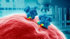
Taking on Cancer Cells Too?
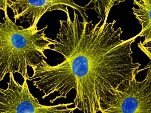
But wait, there’s more! Beyond bacteria, the researchers also tested the MOAg-NPs against cancer cells. They used HeLa cells, a common type of human cervical cancer cell line used in labs. They also tested the *Moringa* extract by itself to see if the plant extract alone had an effect.
Guess what? The *Moringa* extract on its own didn’t really do much to inhibit the growth of the HeLa cells, even at high concentrations. But the MOAg-NPs? Totally different story! They showed strong growth-inhibitory properties against the cancer cells. The effect was dependent on both the concentration of nanoparticles and how long the cells were exposed. Even at the lowest concentration tested, the nanoparticles significantly inhibited cell growth after 24 and 72 hours. The longer exposure times were much more effective than a short 4-hour exposure.
This is incredibly exciting! It suggests that these green-synthesized nanoparticles derived from hydroponic *Moringa* not only fight bacteria but also have the potential to fight cancer cells. The exact mechanism against cancer cells is still being explored for nanoparticles in general, but it often involves generating reactive oxygen species (ROS), which cause damage to cancer cells, and potentially disrupting mechanisms that cancer cells use to pump out drugs.
The Big Picture and What’s Next
So, what have we learned? This study successfully demonstrated a simple, green, and cost-effective way to make silver nanoparticles using extract from hydroponically grown *Moringa oleifera* leaves. These nanoparticles are tiny (around 10 nm), mostly spherical, and crystalline. Crucially, they show significant potential as:
- Antibacterial agents: Effective against both Gram-positive and Gram-negative bacteria, likely by disrupting membranes and inhibiting key energy-producing enzymes like FOF1-ATPase.
- Cytotoxic agents: Capable of inhibiting the growth of cancer cells (HeLa cells) in a dose- and time-dependent manner.
The fact that these nanoparticles came from hydroponically grown *Moringa* is interesting, especially since they seemed more potent and smaller than Ag-NPs made from soil-grown *Moringa* in another study. This hints that the growing conditions might influence the plant’s chemistry and, consequently, the properties of the nanoparticles synthesized from it.
This research is a fantastic step forward, highlighting the power of combining sustainable plant cultivation (hydroponics) with green nanotechnology. It opens doors for developing new therapies for infections and cancer that are potentially safer, more environmentally friendly, and effective against resistant strains.
Of course, like any good science, this is just one piece of the puzzle. More research is needed to fully understand the detailed mechanisms, test these nanoparticles against a wider range of drug-resistant bacteria and other pathogens (like fungi!), and crucially, evaluate their safety on normal, non-cancerous cells and in living systems (like animal models) before they could ever be used in humans. But the potential is clearly there, and it’s pretty inspiring!
Source: Springer