Jiaogulan’s Secret Weapon? This Leaf Extract is Giving Leukemia Cells a Tough Time!
Hey science fans! I’ve got some super exciting news from the world of medical research that I just had to share. You know how we’re always looking for new ways to tackle tough diseases? Well, scientists have been digging into traditional remedies, and sometimes, they strike gold! Today, I want to talk about a plant called Gynostemma pentaphyllum – you might know it as Jiaogulan or “cheap ginseng” – and how it’s showing some serious promise against a particularly nasty type of cancer: Acute Myeloid Leukemia (AML).
The Big Bad: Understanding AML and its Sidekicks FLT3 e WT1
So, cancer. It’s a word we all dread, and leukemia, a cancer of the blood, is a real tough cookie. AML, specifically, is a type where immature myeloid cells in your bone marrow go a bit haywire, multiplying like crazy and not differentiating properly. It’s one of the leukemias with the lowest survival rates, which is pretty grim, I know.
Now, in many AML cases, there are a couple of molecular culprits that help the disease progress. One is a protein called FLT3 (Feline McDonough Sarcoma-like tyrosine kinase 3). When FLT3 is overexpressed or mutated on AML cells, it’s like putting the foot on the gas pedal for cell growth and survival. This makes it a really attractive target for new therapies. Another player is WT1 (Wilms’ tumor 1), a nuclear protein that acts like a marker for leukemic cell proliferation. High levels of WT1 often mean a poorer prognosis. So, finding something that can shut down or slow down FLT3 and WT1? That would be a game-changer!
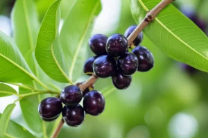
Enter Our Green Champion: Gynostemma pentaphyllum
This is where our leafy hero, Gynostemma pentaphyllum, steps into the spotlight. This climbing vine has been a staple in Traditional Chinese Medicine for centuries, used for everything from common colds to heart disease. Its leaves are packed with bioactive goodies, and it’s often sold as a health supplement or tea.
Scientists already know it contains cool compounds like saponins (especially gypenosides) and flavonoids, which have shown anti-cancer effects against various cancer cell lines. But here’s the kicker: no one had really looked into how G. pentaphyllum might specifically tackle AML by targeting FLT3 and WT1. Until now!
The Scientific Quest: Extracting the Good Stuff
A team of researchers decided to investigate this very question. They got fresh leaves of G. pentaphyllum (a big shoutout to the plantation in Chiang Mai, Thailand, for providing them!), dried them, ground them into a powder, and then got to work with extractions.
They used a method called sequential maceration with three different solvents to get different types of extracts:
- An n-hexane extract (non-polar stuff) – let’s call it F-Hex.
- An ethyl acetate extract (intermediate polarity) – F-EtOAc.
- An ethanol extract (polar stuff) – F-EtOH.
The idea was to see which type of compounds, based on their solubility, would be the most effective. The F-EtOH gave the highest yield, but as we’ll see, yield isn’t everything!

Putting Extracts to the Test: Cytotoxicity Screening
Next up, they tested these extracts against a panel of human leukemia cell lines, including two AML cell lines that are notorious for overexpressing FLT3: EoL-1 (with wild-type FLT3) and MV4-11 (with a mutated, always-on FLT3). They used the MTT assay, a common lab test, to see how much of each extract was needed to inhibit cell growth by 50% (this is called the IC50 value – lower is better!).
And guess what? The ethyl acetate extract (F-EtOAc) was the star! It showed the highest cytotoxicity against both EoL-1 (IC50 = 40.82 µg/mL) and MV4-11 (IC50 = 35.54 µg/mL) cells. The other extracts were much weaker. This was a big clue that the compounds with intermediate polarity were the ones doing the heavy lifting.
Importantly, they also checked the extracts against normal human peripheral blood mononuclear cells (PBMCs). The F-EtOAc extract didn’t show significant toxicity to these healthy cells at the concentrations effective against leukemia cells, which is great news – we want treatments that target cancer cells selectively!
Digging Deeper: Fractionation and Finding F10
Since F-EtOAc was so promising, the researchers decided to break it down further to try and isolate the most active components. They used column chromatography, a technique that separates compounds based on their chemical properties. This process yielded ten main fractions from F-EtOAc.
They tested all these fractions, and one, dubbed F10, really stood out. F10 was even more potent than the original F-EtOAc extract, with IC50 values of 9.68 µg/mL in EoL-1 cells and 16.33 µg/mL in MV4-11 cells. Now we’re talking! According to National Cancer Institute guidelines, an IC50 below 10 µg/mL for a fraction is considered highly cytotoxic.
How Does It Work? Unraveling the Anti-Leukemia Mechanisms
Okay, so F-EtOAc and especially F10 can kill leukemia cells. But how? The scientists dug into the molecular mechanisms:
1. Cell Cycle Arrest: Hitting the Brakes on Cancer Growth
They looked at what the extracts did to the cell cycle – the process cells go through to divide. Using flow cytometry, they found that both F-EtOAc and F10 caused the AML cells to get stuck in the G0/G1 phase of the cell cycle. This means the cells weren’t progressing to division. They also saw an increase in the “sub-G1” population, which is a hallmark of cells undergoing apoptosis (programmed cell death).
2. Apoptosis Induction: Telling Cancer Cells to Self-Destruct
This was a big one! Apoptosis is a clean way for cells to die without causing too much inflammation. The researchers confirmed that F-EtOAc and F10 were indeed pushing the leukemia cells into apoptosis. They used Annexin V/PI staining, which can distinguish between healthy, early apoptotic, late apoptotic, and necrotic cells.
The results were clear: treatment with F-EtOAc and F10, particularly F10, significantly increased the percentage of apoptotic cells in a dose-dependent manner. For instance, F10 at its IC50 concentration ramped up apoptotic cells in EoL-1 to about 24% and in MV4-11 to nearly 19%, compared to much lower levels in untreated cells.

3. Mitochondrial Mayhem: Messing with the Cell’s Powerhouse
Mitochondria, the powerhouses of our cells, play a key role in initiating apoptosis. One sign of mitochondrial distress is a drop in the mitochondrial membrane potential (ΔΨm). The researchers found that F-EtOAc and F10 caused a significant decrease in ΔΨm in the AML cells. This depolarization of the mitochondria is a strong signal that the intrinsic pathway of apoptosis was being triggered.
4. Key Protein Players: p53 and Caspase-3
To get even more specific, they looked at key proteins involved in apoptosis. They found that treatment with F-EtOAc and F10 led to an up-regulation of p53. p53 is a famous tumor suppressor protein; when it’s activated, it can halt the cell cycle or trigger apoptosis. They also saw an activation of caspase-3. Caspases are like the executioners of apoptosis – caspase-3 is a key one that chops up cellular components, leading to cell death. The increase in p53 and active caspase-3 strongly supported the apoptosis findings.
5. Targeting the Bad Guys: FLT3 and WT1 Expression
And what about those AML markers we talked about, FLT3 and WT1? Using Western blotting, the researchers showed that both F-EtOAc and F10 significantly reduced the expression levels of both FLT3 and WT1 proteins in the EoL-1 and MV4-11 cells. This is huge! It suggests that the extract and its active fraction are hitting these critical proliferation and survival pathways in AML cells.
The Star of the Show: Identifying Dehydrovomifoliol
After all this exciting work showing what F10 does, the next big question was: what’s in it that’s so powerful? Through a meticulous process of further fractionation and purification (using techniques like MPLC and RP-HPLC), they isolated a major bioactive compound from F10. Using ESI-MS and NMR spectroscopy (fancy tools for figuring out chemical structures), they identified this compound as dehydrovomifoliol.
Dehydrovomifoliol is a type of compound called a sesquiterpene or megastigmane glycoside. It’s not entirely new to science; it’s been found in other plants, including some in the same family as G. pentaphyllum (Cucurbitaceae), and even in rice! Previous studies on dehydrovomifoliol from other sources have shown it can have cytotoxic effects against other types of cancer cells.
The researchers even did some cool in silico (computer-based) analysis using network pharmacology. This predicted that dehydrovomifoliol could interact with several gene targets crucial in leukemia, like TNF, AKT1, IL6, and CASP3 (caspase-3, which they already saw activated!). The predicted pathways involved signal transduction, cell proliferation, apoptosis, and cell cycle progression – all lining up beautifully with their experimental findings!
Why This is So Cool and What’s Next
I find this research incredibly promising! It shows that an ethyl acetate extract of Gynostemma pentaphyllum leaves, and particularly its fraction F10, has significant anti-leukemia activity against AML cells that overexpress FLT3. It seems to work by:
- Inhibiting FLT3 and WT1 expression (key drivers of AML).
- Inducing G0/G1 cell cycle arrest (stopping cells from dividing).
- Triggering apoptosis (programmed cell death) via the mitochondrial pathway, involving p53 up-regulation and caspase-3 activation.
And they’ve pinpointed dehydrovomifoliol as a major bioactive compound responsible for these effects. How amazing is it that a traditional medicinal plant holds such potential? Most previous studies on G. pentaphyllum focused on more polar compounds, so this work highlights the importance of exploring less polar fractions too.
Of course, this is still early-stage research. These are findings in cell cultures (in vitro). The next steps would involve more in-depth studies to fully understand the mechanisms, test the efficacy and safety in animal models, and eventually, if all goes well, in human clinical trials. But it’s a fantastic start and provides new insights into how G. pentaphyllum might be used to fight acute myeloid leukemia.
It just goes to show, there are so many natural wonders out there still waiting to be explored for their medicinal secrets. Hats off to the scientists for this meticulous and exciting work! I’ll definitely be keeping an eye out for what they discover next.
Source: Springer