Gut Feeling Gone Wrong: Unpacking the Microbial Mayhem in Chronic Atrophic Gastritis
Hey there, science enthusiasts and curious minds! Ever wonder what’s really going on inside your tummy, especially when it’s not feeling its best? Today, we’re diving deep into the world of chronic atrophic gastritis (CAG), a condition that’s more than just a simple stomach ache. It’s a bit of a hot topic, and for good reason, as it’s considered a precancerous lesion for gastric cancer. Yikes! So, understanding it better is pretty crucial, wouldn’t you say?
Now, modern medicine tells us that chronic gastritis comes in two main flavors: chronic non-atrophic gastritis (NAG) and our main character, CAG. Getting to CAG is usually a long journey, a slow burn under the influence of various troublemakers. Think of it as a progression: normal tummy lining, then NAG, then CAG, which can then lead to something called intestinal metaplasia, and eventually, for some, gastric cancer. It’s a pathway we really want to interrupt!
So, What Exactly IS Chronic Atrophic Gastritis?
In simple terms, CAG is a chronic stomach issue where the glands in your stomach lining decide to shrink or reduce in number. Sometimes, this space gets replaced by fibrous tissue or undergoes changes that make it look more like the intestine (that’s the intestinal metaplasia we mentioned). It’s a condition that often pops up in older folks, partly because these gastric glands can naturally thin out with age. However, and this is a bit concerning, with our modern lifestyles – stress, weird diets, you name it – and better diagnostic tools like gastroscopy, we’re seeing CAG more often, and in younger people too. Not cool, stomach, not cool.
What causes this glandular disappearing act? Well, it’s a mixed bag:
- Age: As we get older, factors that nourish the stomach lining, like gastrin, might decrease, or the stomach lining just doesn’t respond to them as well.
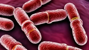
- Helicobacter pylori (HP) infection: This little bacterium is a big culprit. Over 90% of CAG patients have an HP infection. It’s like the unwelcome guest who overstays their welcome and trashes the place.
- Lifestyle habits: Regularly downing strong tea, spirits, coffee, or eating super hot or cold foods can repeatedly damage the stomach lining.
- Medications: Long-term use of high doses of non-steroidal anti-inflammatory drugs (NSAIDs) can mess with the stomach’s natural protective mechanisms.
- Bile reflux: When bile from your intestine flows back into the stomach, it can damage the mucosal barrier.
Current treatments try to tackle these causes – eradicating HP, using acid blockers, and so on. But, these don’t always fundamentally solve the precancerous state, and CAG symptoms can be all over the place, like dull pain, bloating, or nausea, making it tricky to diagnose and treat effectively.
Enter the Micro-Detectives: Microbiome and Metabolome
This is where things get super interesting! With fancy DNA sequencing and bioinformatics, we’ve entered a new era of understanding human microbes. Our digestive system, in particular, is like a bustling city of trillions of microorganisms. This “microbiome” and the chemical substances they produce or modify (the “metabolome”) are crucial for our health. When this delicate balance is off, diseases can arise.
So, a group of researchers decided to put CAG under the microscope, quite literally, by looking at both the bacteria and their metabolic byproducts in the stomach. They took gastric juice samples from 30 CAG patients and 30 NAG patients and went full-on CSI with 16S rRNA sequencing (to see who’s there), metagenomics sequencing (to see what they’re capable of doing), and metabolomics sequencing (to see what chemicals are floating around).

Their goal? To paint a clear picture of what’s different in the stomachs of CAG patients and maybe, just maybe, find some clues about how this disease starts and progresses. It’s like trying to understand why one city (NAG stomach) is thriving while another (CAG stomach) is struggling, by looking at its inhabitants and its economy.
What Did They Find? A Shift in the Gastric Landscape
Okay, let’s get to the juicy bits – the findings! First off, using 16S rRNA sequencing, they found that in atrophic gastritis, the diversity of bacteria decreased. Imagine a vibrant ecosystem suddenly losing some of its species; that’s not usually a good sign, right?
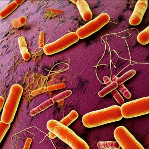
When they looked closer, some bacterial groups were notably different between the CAG and NAG folks. For instance, genera like Prevotella, Rothia, Peptostreptococcus, and Atopobium were more abundant in the NAG (healthier) group. On the flip side, bacteria like Ralstonia, Sphingomonas, Muribaculaceae, and Ruminococcus were more enriched in the CAG group. It’s like different neighborhoods having very different dominant gangs!
The “Most Wanted” Bacteria in CAG
To pinpoint the real key players distinguishing CAG from NAG, the researchers used a clever statistical method called a random forest algorithm. Think of it as asking a super-smart computer to find the most important clues. And who were the prime suspects? The genera that stood out with a significant impact (a “mean decrease in Gini greater than 1.5” for the tech-savvy) included:
- Peptostreptococcus
- Fusobacterium
- Prevotella
- Sphingomonas
- Bacteroides
These weren’t just hanging around; they seemed to be central figures in the microbial drama of CAG.
Metabolic Mayhem: The Chemical Story
It’s not just about which bacteria are there, but also what they’re doing – and that’s where metabolomics comes in. The researchers found 168 metabolites (small molecules involved in metabolism) that were present in different amounts between the CAG and NAG groups. About 81 of these were cranked up in CAG, while 87 were dialed down.
When they looked at what pathways these differentially expressed metabolites were involved in, some interesting patterns emerged. Pathways like:
- Renal cell carcinoma (yes, a cancer-related pathway, which is concerning)
- Proximal tubule bicarbonate reclamation (involved in kidney function and acid-base balance)
- Citrate cycle (TCA cycle – a fundamental energy-producing pathway)
- Aldosterone synthesis and secretion (hormone involved in blood pressure and electrolyte balance)
were significantly enriched with these differential metabolites. This suggests that CAG isn’t just a local stomach problem; it’s messing with some pretty fundamental bodily processes.

Connecting the Dots: Bacteria and Their Chemical Fingerprints
Now for the really cool part: connecting the bacterial changes with the metabolic changes. The study constructed a network showing how the different bacteria and different metabolites were correlated. And guess what? Those “most wanted” bacteria – Peptostreptococcus, Fusobacterium, Prevotella, and Sphingomonas – were found to be in pivotal positions in this network, linked to many different metabolites. It’s like finding out the gang leaders are also controlling various black markets (the altered metabolic pathways).
This strongly suggests that these specific bacteria aren’t just bystanders; they might be actively causing trouble by altering various metabolic pathways, contributing to the development of CAG.
A Deeper Dive with Metagenomics: What Are These Bugs DOING?
To get an even more detailed picture, especially about the functional capabilities of these microbes, the team used metagenomic sequencing. This method looks at all the genetic material in the sample, not just a marker gene like 16S rRNA. Interestingly, while the overall diversity didn’t show a huge statistical difference with this method (sometimes different techniques give slightly different views of the same complex picture), it did highlight some important functional shifts.
The metagenomic data reinforced some of the 16S findings, showing significant differences in genera like Peptostreptococcus, Fusobacterium, Prevotella, and Bacteroides. But it also shone a light on specific species and, crucially, on the functions these microbial communities were performing differently in CAG.
Functional enrichment analysis (using the KEGG database, a go-to for this stuff) revealed that certain metabolic pathways were significantly altered. In the CAG group, pathways like:
- Viral carcinogenesis (again, a cancer link – yikes!)
- Glycine, serine, and threonine metabolism (amino acid pathways)
- RNA polymerase (essential for gene expression)
- Galactose metabolism (sugar metabolism)
- Retinol metabolism (Vitamin A pathway)
were enriched. This means the microbial community in CAG stomachs might be geared towards activities that could, unfortunately, promote disease progression.

What Does This All Mean for Us?
This study is pretty exciting because it’s one of the first to use this triple-whammy approach (16S rRNA, metagenomics, and metabolomics) on gastric fluid from CAG patients. It paints a much richer picture than looking at just one aspect.
The big takeaway? The bacterial community (our microflora) and their chemical byproducts (metabolites) are seriously out of whack in chronic atrophic gastritis. It’s not just about H. pylori anymore; it’s a whole ecosystem shift. The study highlights that bacteria like Peptostreptococcus, Fusobacterium, Prevotella, Sphingomonas, and Bacteroides seem to be key distinguishing features of CAG. They’re not just different; they appear to be actively involved in the disease by messing with important metabolic pathways, including the citrate cycle and even pathways linked to cancer development like renal cell carcinoma and viral carcinogenesis.
Think of our gut microbes as tiny chemists. In a healthy stomach, they’re doing good work. But in CAG, it seems some of the “rogue” chemists take over, producing and altering substances that contribute to the unhealthy state of the stomach lining and potentially pave the way for more serious issues.
Of course, every study has its limitations. The sample size here was relatively small (30 CAG vs. 30 NAG), and it didn’t break down results by age or sex, which can also influence our microbes and metabolites. Plus, these are associations – we still need more research, including lab experiments, to confirm that these specific bacteria cause these changes and to understand the exact biological nitty-gritty.
The Bottom Line
So, what’s the final word? Well, it seems that Peptostreptococcus, Fusobacterium, Prevotella, Sphingomonas, and Bacteroides are more than just names on a list; they’re emerging as essential features that help us tell CAG apart from NAG. And they’re not just passively sitting there – they seem to be mucking up various metabolic pathways.
Furthermore, the enrichment of pathways like viral carcinogenesis, and those involved in amino acid, RNA, sugar, and vitamin A metabolism in CAG, hints at complex mechanisms that could be driving the occurrence and progression of this condition. This kind of research is super important because it could lead to new ways to diagnose CAG earlier, or even new treatments that target these microbial and metabolic imbalances. Imagine being able to rebalance your stomach’s “ecosystem” to help prevent or treat CAG! That’s the hope, anyway.
It’s a fascinating, complex world inside our stomachs, and we’re only just beginning to understand the intricate dance between our cells, our microbes, and our health. Stay curious!
Source: Springer