Spotting Trouble Early: How Super-Resolution Imaging is a Game-Changer for Follicular Lymphoma
Hey there! Let’s dive into something pretty important in the world of lymphoma. You know, follicular lymphoma (FL) is often seen as the ‘chill’ kind of lymphoma – it’s usually slow-growing and most folks do quite well with it for a long time. We’re talking a 5-year survival rate that’s really encouraging, up to 90.6%! But, and this is a *big* but, it has this tricky habit. For about 15–19% of patients, over several years, it can decide to transform into something much more aggressive, called diffuse large B-cell lymphoma (DLBCL). This transformation, or HT as we call it in the medical world, really throws a wrench in things and significantly worsens outcomes.
The Follicular Lymphoma Puzzle
Think of FL like a quiet neighbor; usually keeps to itself, doesn’t cause much fuss. But sometimes, that neighbor can suddenly become very disruptive. That’s the transformation we’re talking about. It happens at an estimated annual rate of 2–3%. The challenge? We haven’t had a super reliable way to predict *who* this will happen to or catch it early. And trying to preemptively blast everyone with chemotherapy isn’t the answer either, as it can actually make things worse for those who *do* transform later. So, catching this transformation early is absolutely critical. It means we can step up the treatment game right when it’s needed, potentially improving how patients fare.
The Challenge of Transformation
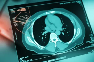
Right now, confirming this HT requires a biopsy. You need a piece of tissue to look at under the microscope. The problem is, FL can be spatially heterogeneous – meaning the transformation might only be happening in one spot, not everywhere. This often means needing multiple biopsies from different sites, which is invasive and, let’s be honest, nobody wants more biopsies than necessary!
We’ve tried using imaging like FDG-PET scans. These scans can show areas of high metabolic activity, which *can* indicate aggressive lymphoma. Higher SUV (standardized uptake value) numbers *can* suggest aggression. But here’s the rub: there’s overlap. Some indolent lymphomas can have high SUVs, and some transformed ones might not have sky-high numbers. This overlap makes PET-CT a bit controversial for *confirming* HT specifically. Plus, PET scans involve radiation and they’re not exactly cheap, limiting how often we can use them just for monitoring. We really needed something better – something non-ionizing, cost-effective, and highly specific to pinpoint those risky areas and guide biopsies more accurately.
Why Blood Vessels Matter
Okay, let’s get a little sciency, but I promise to keep it charming! Researchers have been looking closely at the microenvironment within lymphomas, especially the tiny blood vessels. It turns out these microvessels play a role in how FL progresses. Things like tumor-associated macrophages (TAMs) hanging around can promote new vessel growth and remodeling. The absence of certain types of vessels is also linked to aggressive lymphoma. This got us thinking: maybe changes in the microvascular structure – like how dense the vessels are or how they’re arranged – could be valuable clues for detecting HT.
Enter Super-Resolution Imaging
This is where some seriously cool technology comes in: super-resolution microcirculation imaging. We’re talking about techniques like Maximum Intensity Projection (MIP) and Ultrasound Localization Microscopy (ULM). These aren’t your grandma’s ultrasound! They give us incredibly detailed views of those tiny blood vessel networks. MIP is great for capturing how blood flows through the area over time, sort of like watching a dynamic map. ULM is even fancier; it tracks tiny microbubbles in the bloodstream to map out the microvessels at a really high resolution.

So, the big idea behind this study was to see if these super-resolution techniques could help us spot those high-risk HT regions, guide biopsies right to where the action is, avoid missing the diagnosis, and cut down on those unnecessary repeat procedures.
Putting the Tech to the Test
The study had two parts, kind of like a detective story. First, a retrospective look back (from 2018 to 2022) at patients with either indolent FL or aggressive DLBCL. The goal here was to see if we could find imaging markers that consistently showed up in the aggressive cases. Then, the prospective part (from 2022 to 2024) enrolled new FL patients to see if those markers we found in the first phase actually worked in real-time to differentiate aggressive transformed FL (tFL) from regular FL. Ethical approval? Check. Patient consent? Check. It was all done by the book.
What We Learned First
In that initial look back at 52 patients, we saw some interesting things with conventional ultrasound and Color Doppler. Aggressive DLBCL patients were more likely to have lymph nodes where the normal ‘hilum’ (the indentation where vessels enter) was absent. And the blood flow patterns were different – FL often had a nice, central ‘hilar flow’, while DLBCL was more likely to show a ‘mixed flow’ from multiple sources. This ‘mixed flow’ became a suspicious sign for screening.
However, when we looked at quantitative measures from standard contrast-enhanced ultrasound (CEUS), like peak intensity (which reflects vessel density), there wasn’t a significant difference between FL and DLBCL. This was a key finding: maybe it’s not just *how many* vessels there are, but *how they’re arranged* that matters.
This is where MIP started to shine, even in a small subset of patients with high-quality images. FL often showed a “firework” pattern – like a single point exploding outwards. DLBCL, on the other hand, had a “starry-sky” pattern – lots of scattered dots appearing over time. This really suggested that changes in vascular *structure* were important clues for transformation.
The Main Event: Validation
Now, onto the prospective study with 80 new FL patients. This is where we really tested those potential indicators. We used Color Doppler and, if needed, PET SUVmax to pick suspicious peripheral lymph nodes for further imaging (CEUS, MIP, ULM) and biopsy.
What did we see? Regular FL patients typically showed that nice hilar flow on Doppler, centrifugal enhancement on CEUS, the “firework” pattern on MIP, and classic central vessels on ULM.
But for the patients whose FL had transformed (tFL), the super-resolution images told a different story. On MIP, they showed “mixed enhancement” patterns – either that “starry-sky” look or multiple “firework” origins from different spots. ULM images backed this up, showing chaotic vessel orientations or multiple flow sources.

The Numbers Speak for Themselves
So, how did the new tech perform? Pretty darn well! When we combined MIP and ULM, they showed impressive diagnostic accuracy (97.5%) for identifying HT. Get this: they had excellent sensitivity (100%) and specificity (97.14%). That means they were great at finding the HT when it was there and great at *not* saying it was there when it wasn’t.
Compared to LDH levels (a blood test) and even PET SUVmax (>10 threshold), the super-resolution imaging held its own and, in some ways, was better. LDH was specific but not very sensitive (missed cases). SUVmax was sensitive but not very specific (lots of false positives).
Catching What Others Miss
This is a big deal. The study highlighted cases where standard CEUS missed HT, but the super-resolution imaging caught it. Even some cases missed by Color Doppler due to its angle limitations were visible with ULM. It really seems like looking at the *structure* with this advanced tech gives us an edge.
The findings also lined up nicely with what happened to patients after treatment. The MIP-ULM patterns showed strong agreement with the final diagnosis after six cycles of therapy. The “firework” pattern mostly matched FL, and the “mixed enhancement” mostly matched HT. This suggests the imaging isn’t just a snapshot; it’s predictive.
Crucially, the new method had a significantly lower false-positive rate for HT detection (1.54%) compared to PET/CT using the >10 SUVmax threshold (39.53%). Think about that – way fewer times telling someone they *might* have transformation when they don’t. This is huge for reducing unnecessary biopsies!
Why This Matters for Patients
Catching HT early is key to getting the right treatment at the right time. This super-resolution imaging technique looks incredibly promising for identifying those high-risk areas *before* a biopsy. It can guide the biopsy needle right to the suspicious spot, increasing the chances of getting a definitive diagnosis on the first try. This means fewer biopsies, less anxiety, and potentially faster access to the necessary chemotherapy escalation. It really feels like a step towards more precise, patient-friendly diagnostics.
The findings also reinforce the idea that it’s the *remodeling* of the blood vessels, the change in their structure and pattern, that’s the critical indicator of HT, rather than just how many vessels are there. This aligns with other research pointing to the role of things like TAMs in creating this altered vascular architecture.

Compared to current tools like LDH or PET, which can be limited in providing a precise location or suffer from lower specificity, MIP-ULM seems to offer a more nuanced and accurate picture of what’s happening at the microvascular level.
Acknowledging the Hurdles
Now, no study is perfect, and we need to be realistic. Ultrasound, even fancy super-resolution ultrasound, has its limitations. It’s great for peripheral lymph nodes (like in the neck, armpit, groin), but it struggles with deep locations like the mediastinum (chest) or retroperitoneum (abdomen). These deep areas are challenging for biopsies too. The study suggests that if we don’t find evidence of HT in the peripheral nodes using MIP-ULM, then a PET-CT guided biopsy might still be necessary for high-SUV deep lesions.
Also, the number of patients in the prospective study, while sufficient for the main goal, wasn’t huge, so comparing the diagnostic performance *statistically* between *all* the different techniques was tricky. And since it was done at a single center, we need to see these results replicated elsewhere to be completely sure they hold up.
Looking Ahead
Despite the limitations, this pilot study is really exciting! It shows that super-resolution microcirculation imaging using MIP and ULM is a cost-effective and promising tool for detecting HT in FL. It significantly enhanced diagnostic precision, reduced the risk of missing HT, and could potentially avoid unnecessary invasive procedures.
The researchers are already thinking about the future, like developing non-contrast versions of this imaging (even less hassle for patients!) and figuring out the best way to integrate this into routine clinical practice. The goal is to optimize how we manage FL and keep improving patient outcomes.
The Bottom Line
For me, seeing technology like this emerge is incredibly hopeful. It means we’re getting smarter about fighting complex diseases like lymphoma. Being able to spot that critical transformation early, guide treatments more effectively, and potentially reduce the need for multiple invasive tests? That’s not just good science; that’s better care for patients. This super-resolution imaging is definitely a technique to keep our eyes on!
Source: Springer