Peeking Inside the Developing Brain: How DNA Folds Shape Our Minds
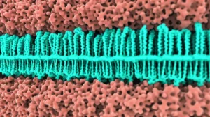
Hey there! Ever wonder how that incredibly complex organ, the human brain, actually builds itself? It’s not just about which genes are turned on or off, but also *how* the DNA is folded up inside the cell nucleus. Think of it like origami – the same piece of paper can become vastly different shapes, and those shapes determine its function. In our cells, this DNA origami, called chromatin, folds into intricate 3D structures, and these structures play a massive role in deciding which genes get expressed and when.
I recently dove into some really fascinating research that looked at this very thing during a critical time in human development: the fetal stage, specifically from about 11 to 26 weeks gestation. This is when different parts of the brain are really starting to specialize and take shape. The big question was: how does the 3D organization of chromatin change across different brain regions and over time, and what does that mean for development and even brain disorders?
Mapping the Fetal Brain’s 3D Landscape
So, the researchers behind this study (and by ‘I’ and ‘we’ from now on, I’m often referring to the collective effort and findings presented in this paper, like I was part of the team uncovering these secrets!) decided to create a detailed map, an “atlas” if you will, of the 3D chromatin structure across six key developing human brain regions. We’re talking about:
- The Prefrontal Cortex (PFC): The brain’s executive suite, involved in complex thought and decision-making.
- The Primary Visual Cortex (V1): Where the brain first processes what your eyes see.
- The Cerebellum (CB): Important for coordination and balance.
- The Corpus Striatum (CS): A subcortical area involved in movement and reward.
- The Thalamus (TL): A relay station for sensory information.
- The Hippocampus (HP): Crucial for memory formation.
Getting samples from these specific regions during this precise developmental window is no small feat, but it was essential for understanding how they become distinct. We used some pretty advanced techniques like in-situ Hi-C to capture the 3D contacts between different parts of the DNA, RNA-seq to see which genes were active, and ChIP-seq to map important regulatory elements like CTCF binding sites and H3K27ac marks (which often highlight active enhancers).
What we found right off the bat was pretty neat: each of these brain regions developed its own unique 3D chromatin signature, and these signatures changed significantly as development progressed. It turns out, this spatiotemporal (that’s just fancy talk for ‘space and time’) dynamic organization of chromatin is a key player in regulating how each brain region develops its specific functions.

The Dynamic World of A/B Compartments
One of the fundamental ways chromatin is organized is into A and B compartments. Think of the A compartment as the ‘active’ zone, full of genes that are being expressed, and the B compartment as the ‘inactive’ or ‘silenced’ zone. We saw that these compartments weren’t static; they were incredibly dynamic during fetal development.
Comparing the developing brain regions to earlier stages like the human blastocyst (a very early embryo) and stem cells, we saw clear differences. What was really cool was how these compartment changes correlated with gene expression. When a region of DNA switched from the inactive B compartment to the active A compartment, the genes within that region tended to get turned up. Conversely, switching from A to B meant genes were likely turned down.
For example, in the Thalamus, genes that switched to the A compartment during development were enriched in functions related to hypothalamus development and glutamate receptor signaling – stuff you’d expect to be important for a sensory relay station! In the Prefrontal Cortex, genes switching to A were involved in forebrain development, axon guidance, and synapse assembly. This tells us that these large-scale structural changes in chromatin are tightly linked to the specific jobs each brain region needs to do as it develops.
TADs: The Structural Neighborhoods
Zooming in a bit, chromatin is also organized into ‘Topologically Associating Domains’ or TADs. These are like insulated neighborhoods where DNA within a domain interacts frequently, but interactions *across* domain boundaries are much less common. TADs help ensure that enhancers (DNA elements that boost gene expression) only talk to the right genes within their neighborhood.
We looked at how these TAD structures differed across the brain regions. Using a technique called PCA (Principal Component Analysis), which helps visualize differences, we saw that regions like the PFC, V1, and Hippocampus tended to cluster together, while the Cerebellum and Thalamus were more distinct. This makes sense, as the PFC, V1, and HP are all part of the forebrain’s cerebral cortex or related structures, while the CB is hindbrain and the TL is diencephalon.
What was particularly interesting was the divergence between the PFC and V1. Even though they’re both cortical areas, they handle very different jobs (complex thought vs. vision). At earlier fetal stages (GW11-14), their TAD structures were quite similar. But around GW16, they started showing clear differences, which became even more pronounced later on. This structural divergence coincided with differences in gene expression patterns related to their specific functions – PFC genes involved in neural precursor cell proliferation, and V1 genes involved in synaptic signaling and assembly. This suggests that changes in TAD organization contribute to the functional specialization of these areas.
Chromatin Loops: Connecting the Dots
Even finer than TADs are chromatin loops. These are direct physical contacts between distant pieces of DNA, often bringing an enhancer element into close proximity with the promoter of the gene it regulates. These loops are absolutely critical for fine-tuning gene expression and establishing cell identity.
We identified hundreds of thousands of these loops across the different brain regions and developmental stages. Just like the compartments and TADs, these loops showed remarkable region and stage specificity. Loops present in the Thalamus might be absent in the Cerebellum, and loops present early in development might disappear later on.
Genes linked by these region-specific loops were often involved in functions specific to that brain area. For instance, loops specific to the Thalamus at GW16 were linked to genes involved in synapse organization, including LHX9, a known regulator of thalamic neuron development. We also saw loops specific to the telencephalon regions (PFC, V1, CS) around genes like FOXG1, which is important for establishing cortical subdivisions.
Beyond region-specific loops, we also found loops that were specific to certain developmental stages. Early-stage loops (GW11/13) were linked to genes involved in cell proliferation and basic cellular processes, while late-stage loops (GW23/26) were associated with genes related to nerve branching and signaling pathways. This dynamic looping landscape helps orchestrate the complex sequence of events during brain maturation.
Super-Enhancers: Powering Regional Identity
Among the regulatory elements involved in looping, super-enhancers (SEs) stood out. These are dense clusters of enhancers that are particularly strong drivers of gene expression and are often associated with genes that define cell or tissue identity. We found hundreds of these SEs in the developing brain, many of which were brain region-specific.
These region-specific SEs were often involved in mediating region-specific loops. For example, in the Cerebellum, we found SEs and associated loops linked to genes like ZIC1 and ZIC4, which are highly expressed in the CB and involved in hindbrain development. These loops and SEs were absent in the PFC and V1. Similarly, PFC/V1-specific SEs and loops were found around genes like POU3F3, important for cortical development.
What was really striking was that SEs didn’t just interact with gene promoters; they also seemed to prefer interacting with *other SEs*, sometimes even across long genomic distances that span multiple TADs. This suggests that SEs might form higher-order interaction hubs, creating a powerful regulatory network that reinforces brain region-specific gene expression programs.

Linking Structure to Function: The SLN Story
To really prove that these 3D chromatin interactions were functionally important, we picked a specific example. We looked at the SLN (Sarcolipin) gene, which was highly upregulated in the PFC during later fetal stages (GW23/26) and is known to be expressed in certain neurons. Using our 3D interaction data, we identified a potential enhancer region located about 140 kb away from the SLN gene promoter that strongly interacted with it in the later PFC samples.
To test if this enhancer was actually important, we used CRISPR/Cas9 gene editing to remove this specific DNA sequence in human embryonic stem cells. We then grew these edited cells into cortical organoids – miniature, 3D structures that mimic aspects of the developing human cortex. Comparing the organoids where the SLN enhancer was knocked out to control organoids, we saw a significant reduction in the number of mature neurons. The progenitor cells (the ones that *become* neurons) were still there, but they weren’t maturing properly. This provided direct evidence that this specific enhancer, identified through 3D chromatin interactions, plays a functional role in neuron maturation during human cortical development.
Connecting the Dots to Neuropsychiatric Disorders
Finally, we wanted to see if this 3D chromatin atlas could shed light on brain disorders. Large-scale studies (GWAS) have identified thousands of genetic variations (SNPs) associated with conditions like schizophrenia, autism, ADHD, Alzheimer’s, and others. Most of these SNPs don’t sit directly *in* genes but in the vast non-coding regions between them. This makes it tricky to figure out which gene they actually affect.
By integrating our 3D chromatin interaction data, we could link many of these GWAS SNPs to specific genes, even if they were located far away in linear DNA sequence. We identified hundreds of these “SNP-linked genes” across the different brain regions for various neuropsychiatric disorders and traits like intelligence (IQ).
For example, a SNP associated with schizophrenia was found to interact via a chromatin loop with the LINC00461 gene in the PFC. This gene is mainly expressed in the brain and has been implicated in brain tumors. Another ADHD-associated SNP didn’t link to the closest gene, but instead interacted with the ZNF217 gene in the PFC, which is known to affect cell proliferation – relevant for early brain development.
Looking at the functions of these SNP-linked genes, we found they were often involved in nervous system development processes, providing molecular clues about how these genetic variations might contribute to disease risk. Interestingly, when we compared the genes linked to different disorders, we saw a significant overlap among neurodevelopmental disorders like schizophrenia, autism, and ADHD, suggesting they might share some common underlying genetic pathways. Neurodegenerative disorders like Alzheimer’s and Parkinson’s, however, had more distinct sets of linked genes, pointing to more unique molecular mechanisms.
Wrapping It Up
So, what’s the big takeaway from all this? Well, it’s clear that the 3D folding of our DNA isn’t just some random mess; it’s a highly organized and dynamic system that changes over time and is specific to different parts of the developing brain. This intricate 3D architecture, especially the dynamic A/B compartments, TADs, and chromatin loops mediated by super-enhancers, plays a crucial role in fine-tuning gene expression and guiding the unique development of each brain region.
Plus, having this 3D map helps us make sense of those mysterious genetic variations linked to neuropsychiatric disorders. By connecting SNPs in non-coding regions to their target genes via chromatin interactions, we get a much clearer picture of the potential molecular players involved in these complex conditions.
Of course, this study used bulk tissue samples, meaning we saw the average 3D structure across all the different cell types in that region (neurons, glial cells, etc.). Future research using exciting new single-cell technologies will be needed to really tease out the 3D chromatin landscape in specific cell types within the developing brain. But for now, this atlas provides a fantastic resource and some seriously cool insights into how the brain’s 3D genetic blueprint unfolds to create the incredible organ that makes us, well, us!
Source: Springer