Fixing Stubborn Breaks: How a Little Piece of Your Knee Can Heal Your Thigh Bone
Hey there! Let me tell you about something pretty remarkable happening in the world of fixing broken bones, especially those really stubborn ones that just refuse to heal. You know, sometimes a fracture doesn’t quite get the memo about knitting itself back together. We call that a nonunion, and when it happens in the distal half of your femur – that’s the lower part of your big thigh bone, near the knee – it can be a real headache.
Why is it so tough? Well, often, these areas don’t have the best blood supply, and they might be missing those crucial bone-building cells needed for healing. Traditional bone grafts, where we take bone from one place and put it at the break, don’t always work because they might not bring enough life (vascularity) to the party. So, we need something with a bit more punch.
The Challenge of Nonunion
Imagine you’ve broken your femur, and despite trying to fix it, it just won’t heal. This “recalcitrant” nonunion, especially after previous attempts like using nonvascularized bone grafts, is a significant hurdle. When the bone ends are “atrophic,” meaning they’ve kind of withered away without good blood flow, they lack the biological spark to bridge the gap.
Surgeons have several tricks up their sleeves for these tough cases, like the Masquelet technique, using vascularized bone flaps (bone with its blood vessels attached), or external fixators like the Ilizarov method. For big gaps (more than 5 cm), a vascularized fibular flap (taking a piece of your smaller lower leg bone) is a go-to. But for smaller gaps, say less than 3 cm, the fibular flap can be tricky. Finding the right spot for the nutrient artery can be hard, and sometimes you have to trim the fibula piece, potentially messing with its blood supply.
Enter the Medial Femoral Condyle Flap
This is where our star player comes in: the medial femoral condyle (MFC) corticoperiosteal flap. Think of it as a small, living patch of bone and its covering (periosteum) taken from the inside part of your knee area. What makes it special is that it comes with its own dedicated blood vessels, usually the descending genicular vessels, which branch off the main femoral artery. These vessels are like a lifeline, ensuring the flap is *vascularized* – full of life and ready to work.
This flap is particularly useful for nonunions in the distal half of the femur with minimal or small gaps (up to 3 cm). Why? Because it brings those essential osteogenic cells (the ones that build bone) and growth factors directly to the nonunion site. It’s like delivering a biological healing kit right where it’s needed most.
But here’s the crucial part: no matter how good the biological stuff is, the mechanics have to be right. Adequate rigid fixation is absolutely paramount. You need to stabilize the break so there’s minimal movement (less than 10% strain, if you want to get technical, according to Perren’s strain theory). Combining this mechanical stability with a biologically active flap is the winning formula for atrophic nonunions.
The beauty of the pedicled MFC flap for distal femur nonunion is that it can be harvested relatively easily from the lower part of the medial femur and rotated upwards to cover the break after the fracture is stabilized. It’s like swinging a gate shut over the gap.
Our Little Study
We wanted to see just how well this pedicled MFC corticoperiosteal flap works for those really tough, “recalcitrant” nonunions in the distal half of the femur. We also wanted to keep an eye out for any complications.
So, we gathered a small group – just three male patients – who had these stubborn nonunions in the distal half of their femur. Their average age was about 36.7 years. These weren’t easy cases; they all had recalcitrant nonunion, meaning previous treatments hadn’t worked. Two had aseptic (non-infected) atrophic nonunions, and one had a history of septic (infected) nonunion. They had been dealing with this for a long time, averaging 60 months of nonunion, and had already had multiple surgeries (two to three each). The gaps at the nonunion site were generally small, around 10 mm, though one patient initially had a larger defect that had been managed differently before.
Before surgery, we did a little measurement check called the transposition ratio. This helps us figure out if the flap, with its blood supply attached, can comfortably reach the nonunion site without being stretched too tight. We measured the distance from the medial femoral epicondyle (a bony bump near your knee) to the nonunion site and divided it by the distance from the epicondyle to the lesser trochanter (another bony landmark higher up the femur). If this ratio was too high (we set a limit of 0.5, though others have used up to 0.6), the flap might not reach reliably. Luckily, all our patients were within the acceptable range.

The Procedure
For the patient with the history of infection, we took a staged approach. First, we cleaned out the infection, removed old hardware, and put in an antibiotic-loaded cement spacer. Once the infection was under control, we went back in. For all three patients, the core procedure involved:
- Removing any old implants.
- Cleaning up the bone ends until we saw fresh bleeding (“paprika sign”).
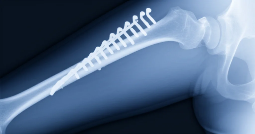
- Putting in new, rigid fixation (like an interlocking nail or a locking plate) to stabilize everything.
- Filling any remaining gaps with an iliac crest bone graft (taken from the hip).
- Harvesting the pedicled MFC corticoperiosteal flap. This involves carefully dissecting down to find those descending genicular vessels and taking a small piece of bone cortex along with the periosteum to keep the vital cambium layer intact.
- Rotating the flap upwards to cover the nonunion site and stitching it in place.
We didn’t use a tourniquet during the flap part of the surgery to make sure we could see the blood flow and confirm the flap was healthy. After closing up, a drain was placed, and a brace was applied to protect the repair.
The Results: A 100% Success Story (So Far!)
Okay, drumroll please… the results were fantastic! All three patients achieved bony union. That’s a 100% union rate in this small series. The average time it took for the bone to heal was about 6.7 months. And importantly, we didn’t see any mechanical failures of the hardware.
Of course, surgery isn’t without its potential hiccups. We did see a couple of complications:
- One patient had a saphenous nerve injury (a nerve near the knee that can cause numbness or tingling) and developed a seroma (a collection of fluid). These often improve over time.
- Another patient developed an incisional hernia at the iliac crest donor site (where we took the hip bone graft from).
Overall, the donor site morbidity (problems where the flap was taken from) seemed minimal, which is a big plus for this technique compared to some others.
We also checked in on how their knees were doing using the Lysholm Knee Score, since the flap comes from that area. At the one-year follow-up, the average score was a very respectable 92 out of 100, suggesting good knee function.
Patient Stories
Let’s quickly look at a couple of the cases to give you a better picture.
There was a 35-year-old guy with an infected nonunion in his femur that had been going on for 30 months, even after trying different things including an external fixator. We had to do a staged approach because of the infection. After clearing the infection and stabilizing the bone, we used the pedicled MFC flap. Union was achieved after six months, which is pretty great for such a long-standing infected case! He did have a bit of trouble later with the nail needing removal due to a recurring sinus, but the bone itself had healed.
Another patient, a 43-year-old who had polio affecting his legs, had a stubborn atrophic nonunion near his knee for 42 months, with pain and broken hardware. He had a 10 mm gap. We removed the old hardware, put in new plates (a main one and an extra medial one for strength), filled the gap with hip bone graft, and added the pedicled MFC flap to the front-inside part of the femur. He achieved union at six months, and it looked solid at the one-year mark.
Putting It All Together
Managing these tough distal femur nonunions is genuinely challenging. They’re biologically compromised, often messed with before, and surgically complex. Our little study, though small, adds to the evidence that the pedicled MFC corticoperiosteal flap is a feasible and effective option, especially when other methods have failed. Getting that 100% union rate in these difficult cases, with a reasonable healing time and no hardware failure, is really encouraging.
This technique works because it directly addresses the core problems: poor blood supply and lack of bone-building cells. By bringing in a vascularized flap, you’re essentially jump-starting the healing process. This is particularly valuable for those “atrophic” bone ends that are just sitting there doing nothing.
It’s important to remember the limitations of this flap. It can only reach so far. In our experience, it reliably reached the midshaft of the femur but not much higher. That’s why calculating that transposition ratio beforehand is key – you need to make sure the flap can actually cover the nonunion site without tension on its lifeline vessels.
The blood supply, usually the descending genicular artery, is pretty consistent, though variations exist. Taking that thin chip of cortex with the periosteum is crucial for preserving the cambium layer, which is rich in those vital osteogenic cells.
Our findings echo what others have reported. Studies by Yoshida, Guzzini, Choudry, Hamada, Ozdemir, Dekeyser, and Dhingra have all shown good success rates with this flap for femoral nonunions, highlighting its effectiveness, relative ease of harvest (compared to free flaps needing microvascular anastomosis), and generally low donor site issues. It’s a great option for surgeons who might not have microsurgical expertise.
Complications are usually minor, like temporary nerve issues or fluid collections. Serious problems like femur fractures at the donor site are rare, especially if you’re careful not to take too much bone from that area (we used hip graft instead of knee graft for filling gaps to minimize this risk).

Looking Ahead
So, what’s the takeaway? For recalcitrant nonunions in the distal half of the femur, the pedicled MFC corticoperiosteal flap looks like a really promising tool in our surgical kit. It successfully brought these three tough cases to union.
However, and this is a big *however*, this was a very small study. Three patients isn’t enough to make sweeping conclusions. We need larger studies, maybe even controlled trials, to really confirm how effective and safe this technique is in a broader population and over the long term. Our follow-up was only one year, so we can’t say much about late issues like hardware problems or re-breaks. We also didn’t use fancy standardized scores to measure things like pain and function, which would be helpful.
There’s also a learning curve involved in harvesting this flap, and dealing with those anatomical variations in blood vessels requires careful dissection. And while donor site problems were minimal here, the potential for rare but serious complications means patient selection and meticulous technique are absolutely critical.
Despite these limitations, our preliminary results are encouraging. This technique offers a valuable option for complex distal femur nonunions, especially when other methods have failed or free flaps aren’t suitable. It shows the power of bringing biological vitality back to a stalled healing process. It’s a neat trick, and one we hope to see used successfully for more patients in the future!
Source: Springer