
Unseen Threats: What We’re Missing in Advanced Colon Cancer Surgery Specimens
Hey there! Let’s chat about something super important in the world of cancer surgery, specifically when we’re dealing with locally advanced colon cancer. You know, the kind that’s gone a bit beyond just chilling inside the colon wall. You’d think that taking out a colon cancer tumor would be pretty straightforward for pathologists and surgeons, right? Most of the time, sure. But when the cancer gets a bit more aggressive, things get complicated. And turns out, we might be missing some critical details that really matter for patients.
We recently took a deep dive into surgical specimens from a big study called the COLOPEC trial. This trial looked at patients with locally advanced or perforated colon cancer. Our mission? To go back and re-examine the pathology slides with a fine-tooth comb, focusing on two specific things: the edges of the tissue that was removed (the surgical margins) and whether there were any tiny spots of cancer already hiding on the lining of the abdomen (synchronous locoregional peritoneal metastases, or SL-PM).
What we found was pretty eye-opening, and honestly, it highlights some areas where surgeons and pathologists need to work even more closely together. Because missing these details isn’t just a paperwork issue; it has real consequences for how likely the cancer is to come back, especially in the abdomen.
The Sneaky Culprits: Margins and Metastases
So, what exactly are we talking about missing? Two main things:
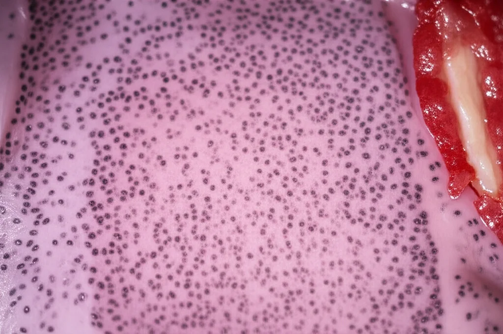
- Positive Resection Margins (R+): This is when, after the surgeon removes the tumor, the pathologist looks at the very edge of the removed tissue under the microscope and finds cancer cells right there. It means the surgeon might not have gotten *all* of the cancer out. In most colon cancer cases, the tumor stays within the bowel wall and the fatty tissue nearby. But in advanced cases, it can grow outwards. The edge we’re worried about isn’t the surface covered by the peritoneum (the abdominal lining), but the edge where the surgeon cut through tissue that *wasn’t* covered by peritoneum – often called the radial margin. Finding cancer here is a big deal.
- Synchronous Locoregional Peritoneal Metastases (SL-PM): This sounds complicated, but think of it as tiny satellite tumors that have already started growing on the abdominal lining (the peritoneum) near the main tumor, *at the same time* the main tumor is found. The TNM staging system classifies these as distant metastases (M1), which means the cancer is more widespread than just locally advanced. This is very different from cancer spreading to nearby lymph nodes (pN), which is considered regional spread.
Why are these tricky? Well, the source text mentions that assessing standard colon cancer specimens is often seen as easy. But for *locally advanced* cases, where the tumor might be stuck to other things or has grown through the wall, it gets much more complex. It takes real skill and attention to detail to properly assess these specimens.
Taking a Second Look: The COLOPEC Reassessment
In the COLOPEC trial, they originally included patients based on imaging showing locally advanced or perforated tumors. For our study, we went back and got the actual pathology slides from 199 of these patients. One pathologist, who’s got a special interest in advanced colorectal cancer, reviewed *all* the slides again, without knowing what the original report said (except for the initial description of the specimen before slicing it up). We also looked at the surgical reports to understand what happened during the operation.
And wow, the differences were significant! According to the original reports, only 5 patients (2.5%) had positive margins (R+). But when we looked again? We found R+ in a whopping 28 patients (14.1%)! That’s a big jump.
It was a similar story for SL-PM. We found these tiny peritoneal metastases in 11 patients (5.5%). But guess how many of those were correctly identified and reported as M1 in the original pathology reports? Just two! The other nine were either missed entirely or misclassified as something else, like being part of the main tumor (pT4a/b) or even mistaken for positive lymph nodes or tumor deposits in the fatty tissue.

Margins, Margins, Everywhere (and Hard to Spot!)
Let’s dig a bit deeper into those positive margins. We realized that R+ isn’t just one thing. Based on where and how the tumor reached the edge of the specimen, we identified a few types:
- Type 1: At the site where the surgeon had to detach the tumor from something it was stuck to (adhesiolysis). This happened in 8 patients. These margins often looked rough, maybe with some bleeding or crushed tissue – signs of mechanical injury. Interestingly, 6 of these were originally called pT4a (tumor growing through the wall) instead of R+ (tumor at the edge of the *cut*).
- Type 2: At the edge of the fatty tissue removed with the colon (the mesocolic resection plane). This was the most common type, seen in 10 patients. Here, we saw tumor growing right up to the inked or cauterized edge.
- Type 3: At the edge of another organ or structure that had to be removed along with the colon tumor (en bloc resection). This happened in 6 patients. Again, tumor at the inked/cauterized edge.
- Type 4: Due to an injury to the specimen itself, like a tear or laceration, that exposed tumor tissue. This happened in 4 patients. These looked like “clean” defects, not related to adhesions. These were also often misclassified as pT4a or even iatrogenic (surgery-caused) perforation.
See the confusion? Especially Type 1 and Type 4 R+ seem to get mixed up with pT4a. Why does this happen? Part of it is probably lack of awareness and training. The edge covered by peritoneum and the radial margin (the cut edge) can look similar, but they mean very different things. R+ means an incomplete removal, while pT4a describes how far the tumor has grown naturally. Also, pathologists often don’t get enough information from the surgical team about what happened during the operation, like if they had to cut through adhesions.
The Hidden Spots: Synchronous Peritoneal Metastases
Now, about those SL-PM. Remember, these are M1 – distant spread. Finding them changes the whole staging and treatment plan compared to just having positive lymph nodes (pN). But we found 9 out of 11 SL-PM cases were missed or misclassified. How?
- Often, these small spots were submitted to pathology as if they were lymph nodes or just part of the main specimen’s fatty tissue.
- Under the microscope, they were then sometimes mistaken for tumor deposits (TDs) or positive lymph nodes within the fatty tissue. TDs are also in the pN category, so classifying an SL-PM as a TD is still a major understaging error (M1 vs pN).
- In one case, a spot on the appendix was thought to be a second primary tumor there, but our review (and later genetic testing) showed it was an SL-PM from the colon tumor.
- Sometimes, they were just missed entirely or vaguely mentioned without being properly identified and labeled as metastases.
A key issue here is anatomy. Lesions in the greater omentum (a fatty apron-like structure in the abdomen) are *very* likely to be peritoneal metastases because its blood supply isn’t connected to the colon’s drainage system. Recognizing the omentum and examining it carefully is crucial. Again, communication is key. If the surgeon sees something suspicious on the peritoneum during the operation, they need to tell the pathologist clearly, maybe even mark it with a suture or put it in a separate container.
Why This Matters for Patients
Okay, so we’re missing stuff. Big deal? Yes, a very big deal. Our study confirmed that finding R+ margins or SL-PM in the initial specimen is strongly linked to the cancer coming back later in the peritoneum (metachronous peritoneal metastases).
In our analysis, R+ margins were associated with more than double the risk of peritoneal recurrence (Hazard Ratio 2.38). And finding SL-PM? That increased the risk almost sixfold (Hazard Ratio 5.98)! These are independent risk factors, meaning they matter regardless of other things like how deep the tumor grew (pT stage) or how many lymph nodes were involved (pN stage).
Think about it: if an SL-PM is missed and the patient is staged as M0 (no distant spread) instead of M1, they might not get the right follow-up or additional treatments they need to target that peritoneal disease. Similarly, knowing about a positive margin can influence decisions about further treatment or closer monitoring.

So, What Can We Do About It?
The good news is that recognizing these issues points us towards solutions. It’s all about improving quality across the board:
- Better Surgery: Surgeons should try to remove adhesions to the tumor *en bloc* (taking the attached tissue with the tumor) whenever possible, as cancer is often hiding in those adhesions. If adhesiolysis is necessary, clearly communicate this to pathology.
- Better Communication: This is huge. Surgeons need to tell pathologists *everything* relevant about the specimen and the surgery, especially regarding margins and any suspicious spots seen on the peritoneum. Structured pathology request forms could help ensure key information isn’t missed. Pathologists should also feel empowered to contact the surgeon for more details.
- Better Pathology Assessment: Pathologists need specific training on recognizing the different types of R+ margins in complex specimens, including the signs of injury or adhesiolysis sites. They also need to be vigilant about identifying SL-PM, understanding where they commonly occur (like the omentum), and distinguishing them from lymph nodes and tumor deposits based on anatomical location and microscopic features. Reading the surgical report is key!
- Clearer Definitions: The study suggests that even the official staging systems could be clearer, especially regarding the term “perforation,” which is currently used for both natural tumor penetration (pT4a) and surgical injuries (which should often be R+).
The Takeaway
Our deep dive into the COLOPEC specimens showed that positive surgical margins and synchronous peritoneal metastases are significantly underdiagnosed in locally advanced colon cancer cases. These aren’t just academic findings; they are critical pieces of information that predict how the cancer might behave and where it might come back.
Improving the detection of R+ and SL-PM requires a team effort – surgeons and pathologists working together, communicating effectively, and having the right training and awareness. By catching these “unseen threats” early, we can ensure patients get the most accurate staging and the best possible treatment plan, ultimately improving their chances against this challenging disease.
Source: Springer