Brain Signals After DBS: What Blood Tests Reveal
Hey there! Let’s chat about something pretty fascinating happening in the world of brain health, specifically for folks dealing with movement disorders like Parkinson’s Disease, dystonia, or essential tremor who might be considering or have had Deep Brain Stimulation (DBS) surgery.
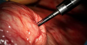
DBS is a fantastic tool, really. It’s like giving your brain a little electrical tune-up to help manage those tricky symptoms. But, like any surgery, especially one involving the brain, there are things we need to keep an eye on. We’re talking about potential risks like swelling around the implanted lead (they call it peri-lead edema, or PLE) or changes in how your brain works, like cognitive decline, after the procedure.
Now, the tricky part is, we don’t fully understand *why* these things happen in everyone, and even tougher, we don’t have a super simple way, like a quick blood test, to predict who might be more at risk or to spot these issues early. Wouldn’t it be great if we did?
That’s where our little adventure into biomarkers comes in. Think of biomarkers as tiny messengers in your blood that can tell us what’s going on inside your body, including your brain. We decided to look at two specific ones after DBS surgery to see if they could give us some clues about what happens to the brain during and after the procedure.
We focused on two proteins:
- Serum glial fibrillary acidic protein (sGFAP): This one is like a report card for glial cells, specifically astrocytes. These cells are part of the brain’s support system, and when they get stressed or injured, sGFAP levels can go up.
- Serum neurofilament light chain (sNfL): This is more about the neurons themselves, the main communication lines in your brain. If the axons (the long parts of neurons) are damaged, sNfL can be released.
So, we followed 58 patients who had DBS surgery at our center for about six months. We took blood samples at different times – before surgery, a few days after, a few weeks after, and then a few months down the line – to see how the levels of sGFAP and sNfL changed.
The Tale of Two Markers: Fast vs. Slow
Okay, here’s where it gets interesting. We saw both sGFAP and sNfL levels increase after surgery, which isn’t totally surprising – surgery is a form of trauma, even if it’s carefully controlled. But *how* they increased and decreased was dramatically different.
sGFAP was the speedy one. Its levels shot up pretty quickly after surgery and then came back down to where they started within just a few weeks. It’s like it was reporting on the immediate, acute reaction in the brain’s support system.
sNfL, on the other hand, was more of a slow-and-steady reporter. Its levels rose more gradually and stayed elevated for months before finally returning to baseline. This is partly because sNfL hangs around in the body for a longer time. So, while it tells us there was neuronal injury from the surgery itself (confirming previous findings), its slow kinetics make it less ideal for spotting *new* or *delayed* problems quickly.
Think of it like this: sGFAP is the breaking news alert about what just happened, while sNfL is the documentary that comes out months later, confirming the event but not useful for real-time updates.

What Influences the Numbers?
We also noticed that some things about the patients themselves seemed to affect the baseline levels of these markers, even before surgery. Things like age and body mass index (BMI) played a role, which makes sense because these factors can influence how these proteins are released or cleared from the body. Older patients and those with lower BMI tended to have higher baseline sNfL, for example.
But the really big deal for us was looking at risk factors for complications. We wanted to see if we could predict who might have more of a brain reaction to the surgery based on their characteristics beforehand.
Linking Markers to Risk and Cognition
This is where sGFAP really shone. We found a strong connection between how well patients performed on a standard cognitive test (the MoCA score) *before* surgery and how much their sGFAP levels increased *after* surgery. Basically, patients who had lower cognitive scores before the procedure showed a bigger spike in sGFAP. This suggests that having some degree of cognitive impairment might make your brain’s glial cells more reactive or vulnerable to the stress of surgery.
This finding is super important because cognitive decline is a concern after DBS for some patients, and PLE (the swelling) has also been linked to cognitive issues. Since sGFAP reflects glial activity, and glial activity is thought to be involved in both the immediate surgical response and potentially delayed issues like PLE and cognitive changes, sGFAP could be a valuable early indicator. It might help us identify patients who need closer monitoring or perhaps influence decisions about who is the best candidate for surgery. Our findings even suggest that there isn’t a sharp cutoff for cognitive safety, but rather a gradual increase in risk with lower cognitive performance, arguing for considering DBS earlier in suitable patients.

Immediate Complications and Motor Subtypes
We also peeked at other things. Interestingly, patients who had immediate complications right after surgery showed a significantly higher increase in sGFAP, but not sNfL. This further supports the idea that sGFAP is better at picking up on acute events. We also saw some hints that different motor subtypes of Parkinson’s (like akinetic-rigid vs. tremor-dominant) might have slightly different patterns in how these markers change, suggesting potential differences in vulnerability, but this needs more investigation.
The DBS Stimulation Itself
And just to be clear on another point: our study, like previous ones, didn’t find any evidence that the electrical stimulation from the DBS device itself causes chronic brain damage, at least not as measured by these biomarkers over the study period. The markers went back to baseline, even after stimulation started. That’s reassuring!
Why GFAP is the Star (for Early Stuff)
So, why is sGFAP getting all this attention?
- It responds *fast* to brain injury from surgery, making it great for spotting early issues.
- Its increase after surgery seems linked to preoperative cognitive performance, potentially flagging patients at higher risk for complications like PLE or cognitive decline.
- It specifically reflects glial damage, which is thought to play a role in the brain’s response to the implanted lead and conditions like PLE.
This means sGFAP could potentially be used as a simple blood test to help assess safety after DBS, especially when brain imaging might not be feasible or clear.
Limitations and What’s Next
Now, no study is perfect, and ours has limitations. It was done at a single center, we had some missing data points, and the number of patients with non-Parkinson’s conditions was small. Plus, while we can *suggest* sGFAP might be useful for detecting delayed complications like PLE or predicting long-term cognitive changes based on its dynamics and correlation with preoperative cognition, we didn’t directly test for PLE with imaging in all patients or follow cognitive function long-term in this specific study.
Wrapping it All Up
In summary, our study showed that sGFAP and sNfL tell different stories about brain injury after DBS surgery. sNfL is a long-term indicator of neuronal damage from the surgery itself, but sGFAP’s rapid response makes it a promising candidate for detecting early injury and assessing risk, particularly in relation to preoperative cognitive function. It gives us a potential window into the brain’s immediate reaction and might help us better understand and manage complications down the line. More research is definitely needed, but it feels like we’re one step closer to having a simple blood test help guide care for DBS patients.
Source: Springer