AI’s Sharp Eye: Revolutionizing PCOS Detection with Super-Res Ultrasound
Hey There! Let’s Talk About PCOS
You know, Poly-Cystic Ovary Syndrome, or PCOS for short, is a really common and often tricky condition that affects so many women during their reproductive years. It’s one of those things that can really mess with your body, often showing up as lots of tiny cysts on the ovaries. Getting a handle on PCOS early is super important because it can have some long-term effects if not managed well.
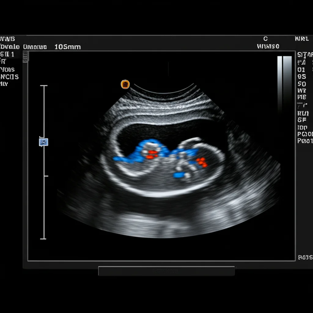
Traditionally, when you suspect PCOS, the doctor usually sends you for an ultrasound. It’s the go-to tool to peek at the ovaries, see their size, shape, and count those little cysts. It’s a vital step, no doubt.
The Challenge with the Human Eye
But here’s the thing: looking at ultrasound images and counting those tiny follicles manually? *Phew*, it can be tough! It takes time, and honestly, what one doctor sees might slightly differ from what another sees. We call that inter-observer variability, and it can make diagnosis a bit less consistent than we’d like. For something as important as PCOS diagnosis, where prompt and accurate results are key to starting the right treatment and preventing potential complications, we really need something more precise and streamlined.
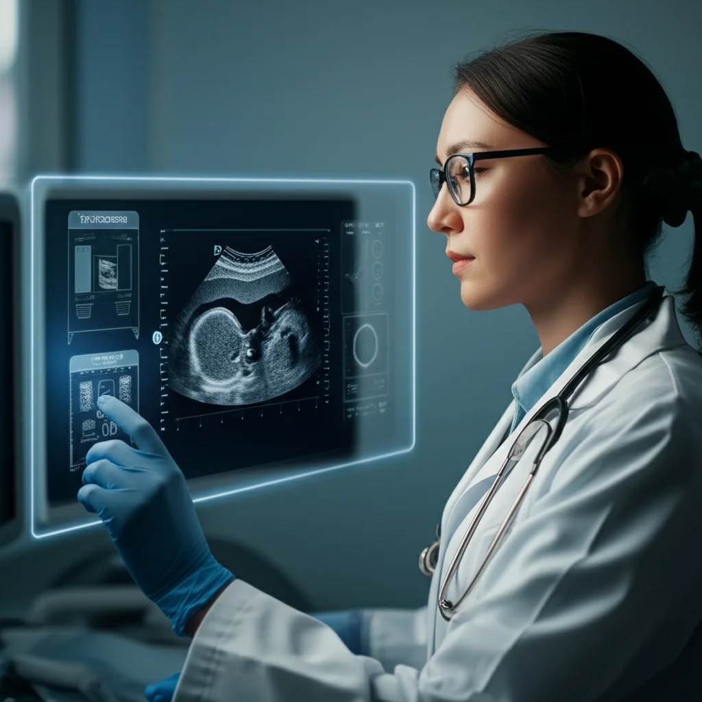
This is where the magic of deep learning comes in! Imagine a super-smart assistant that can look at those ultrasound images and help doctors make a faster, more accurate call. That’s exactly what we’ve been exploring.
Meet QEI-SAM: Our AI Superhero
We’ve developed a cool new integrated approach we’re calling QEI-SAM. It stands for Quality Enhanced Image – Segment Anything Model. The name might sound a bit techy, but the idea is simple: make the ultrasound images better, then use AI to precisely find and outline the important bits (like the cysts), and finally, use another AI to make the diagnosis based on that super-clear info.
We’re leveraging some seriously cutting-edge tech here: Generative Adversarial Networks (GANs) and Convolutional Neural Networks (CNNs). They’re the powerhouses behind getting this system to work like a charm.
Step 1: Making Images Shine with ESRGAN
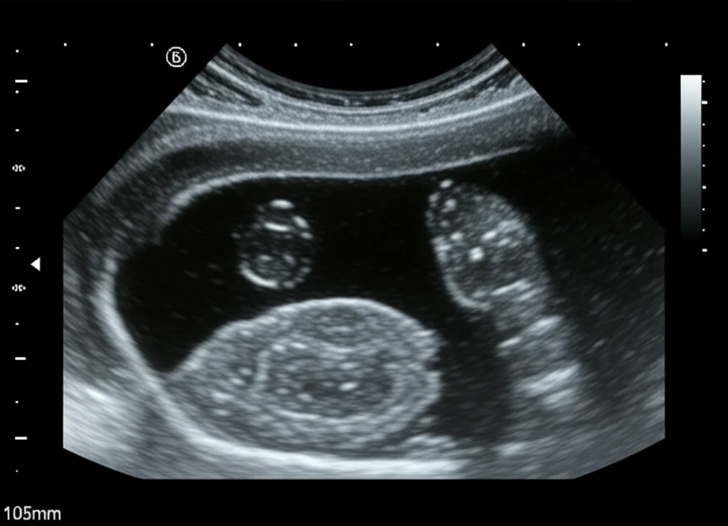
Ultrasound images, while useful, can sometimes lack detail. Think of them like slightly fuzzy photos. To get the best results, we need those images to be crystal clear. Our QEI-SAM model uses something called Enhanced Super Resolution Generative Adversarial Networks (ESRGAN) for this first step.
ESRGAN’s job is to take those standard ultrasound ovary images and give them a serious upgrade. It increases the resolution, sharpens the edges, and brings back the finer structures you might miss in a lower-quality image. We measured its performance using metrics like SSIM, PSNR, and LPIPS (basically, ways to say how good the enhanced image looks compared to a perfect one), and ESRGAN totally outperformed other methods. It achieved an impressive SSIM of 0.938, a PSNR of 38.60, and a low LPIPS of 0.0859. This means we start with the best possible visual information.
Step 2: Spotting the Details with SAM
Once the image is looking sharp, the next crucial step is finding *exactly* where the cysts are. This is where the Segment Anything Model (SAM) comes into play. SAM is pretty revolutionary because it’s designed to segment… well, *anything*! We’ve adapted it specifically for ultrasound ovary images to segment the ovarian cysts.
SAM is like having an incredibly precise digital scalpel that can automatically draw a perfect outline around each cyst. This is vital for accurate diagnosis because things like the number and size of follicles are key criteria. Unlike many older segmentation models that need tons of specific training data for medical images, SAM is much more flexible. It can handle the variations and speckle noise often found in ultrasounds without needing extensive retraining. It just *works*. We evaluated how well SAM segmented the cysts using metrics like the Dice coefficient and IoU score, and it achieved the highest scores compared to other segmentation techniques: a Dice coefficient of 0.9501 and an IoU score of 0.9050. That’s seriously accurate outlining!
Step 3: The Diagnosis with CNNs
With our super-resolution, perfectly segmented images ready, we move to the final step: classification. We feed these processed images into different types of Convolutional Neural Networks (CNNs), which are great at learning patterns in images. We tested several popular ones: ResNet 50, ResNet 101, VGG 16, VGG 19, AlexNet, and Inception v3.
These CNNs look at the enhanced images and the segmented areas to determine if the ovary shows signs of PCOS or if it’s healthy. After putting them through their paces, one model stood out: VGG 19. It achieved the highest accuracy in classifying PCOS from the processed ultrasound images.

Putting It All Together: The QEI-SAM Advantage
The real power comes from combining these steps. When we used the QEI-SAM approach – enhancing the images with ESRGAN and segmenting with SAM *before* feeding them into the CNNs – the results were phenomenal.
Comparing the classification performance *without* QEI-SAM (using just the original images) to *with* QEI-SAM, the improvement was dramatic. Without our enhancement and segmentation, even the best classifier (VGG 19) got around 85.44% accuracy. That’s okay, but not stellar for a medical diagnosis.
But *with* QEI-SAM? VGG 19’s accuracy jumped to an incredible 99.31%! Precision, recall, and F1 score also hit 99%. This is a massive leap and significantly outperforms previous methods tested in similar studies. It statistically confirms that making the images better and precisely identifying the relevant areas makes a huge difference in the AI’s ability to diagnose correctly.
Why This Matters for You (and Everyone)
This isn’t just about cool tech; it’s about making a real difference in healthcare. PCOS diagnosis can be delayed or inconsistent due to the challenges of manual interpretation. Our QEI-SAM approach offers a path to:
- Higher Accuracy: Reducing the chance of misdiagnosis.
- Faster Diagnosis: Streamlining the process for clinicians.
- Reduced Variability: Making the diagnosis more consistent regardless of who is interpreting the image.
- Cost-Effectiveness: Potentially reducing the need for multiple tests or follow-ups due to uncertain results.
By providing physicians with a highly accurate, automated tool based on enhanced, precisely segmented ultrasound images, we can help ensure women get the right diagnosis and start managing their PCOS sooner. It focuses on the visual evidence from the ultrasound, which is often more direct for identifying the key features like cysts compared to just relying on metabolic markers.
The Big Picture
We’re really excited about this QEI-SAM approach. It shows that combining powerful image enhancement (ESRGAN) with flexible, precise segmentation (SAM) and robust classification (VGG 19) can lead to near-perfect accuracy in detecting PCOS from ultrasound images. It’s a significant step towards automating and improving the diagnostic workflow.
Of course, science keeps moving! Future work will involve testing this on even larger and more diverse datasets to make sure it works well for everyone and making the system even more efficient for clinical use. But for now, we’re thrilled with the results and the potential this holds for better PCOS care.
Source: Springer


